The Fetus In Utero
From Mystery to Social MediaImages of the Fetus in Utero
Constructing Images of the Fetus In Utero
Once restricted to the privacy of the doctor’s office, ultrasound images of the fetus are now immediately recognizable in the public arena, through advertising and social media, where posts tagged “baby’s first pic” are commonplace. These depictions of the fetus in utero have become iconic and are arguably the most easily recognized medical image. How and why did this happen? And at what price and to what end?
This exhibition takes an historical approach to this question by exploring the complex evolution of the fetal image in Western Christian culture. We show that before images of the fetus in utero entered the digital age, they have been deployed in three distinctive ways over the past 500 years. First, during the Renaissance, the fetus in utero transformed from an object of divine mystery to one of “rational” inquiry at the hands of male anatomists. Second, from 1700–1965, images of the fetus in uterus underwent a process of medicalization through the male medical gaze. Third, from 1965–present, the fetus in utero has been curated as a public image by a variety of individuals and movements steeped in social and political contexts.
Curated by Brian Callender, MD, Assistant Professor of Medicine, The University of Chicago; and Margaret Carlyle, Postdoctoral Researcher and Instructor, Stevanovich Institute on the Formation of Knowledge, The University of Chicago. This virtual exhibit was created by Teodora Szasz and Ashleigh Cassemere-Stanfield of the Research Computing Center in collaboration with the primary curators.
The Fetal Image & Seeing the World
The story of the image of the fetus in utero told here is one of both historical change and continuity. While the content of the fetal image has changed over time, through new obstetrical and imagining technologies, its function has not. The fetal image has always served as a tool to both reflect and enact certain worldviews. It has never been a value neutral image, but rather exhibits the imprint of social, cultural, and political considerations. The naturalization of the fetal image in religious, medical, political, and entertainment contexts, in turn, has conditioned how we see the world.
In the present-day context, in which reproductive rights are being challenged across America, the time is now more urgent than ever to understand the history of the fetal image. Indeed, the fetal ultrasound’s most powerful and ideologically driven use in our times has been in the context of the abortion debate. In their attempts to limit reproductive rights, the right-to-life movement has extensively used the fetal ultrasound to promote their cause. By presenting and ventriloquizing fetuses, this movement has curated both still and moving images to assert “fetal autonomy” while disembodying the pregnant woman. Such imagery targets the viewer’s emotions in ways to assert “fetal personhood,” while serving as powerful tools in legislative campaigns aimed at restricting women’s reproductive rights.
The fetal image has also achieved iconic status in social media, entertainment, and commerce. In the seemingly more benign context of pregnancy and “gender reveal” announcements, the fetal ultrasound has become a digital badge of impending parenthood, curated with aims of sharing knowledge to a large, but often anonymous, digital social world. Images of the fetus have also been deployed in advertisements to sell cars and snack chips, expanding and further defining how we both utilize and interpret these images.
As this exhibit details, the visual culture of the fetus in utero has a long history that continues to be shaped by the contexts in which these images are produced, consumed, and interpreted. These are to be sure specifically Western contexts and the fetal imagery in other knowledge cultures, from Chinese and Arabic traditions for example, remain to be explored. Moreover, the exhibit reminds us that even the most advanced imaging technologies, although they seem to directly show reality, actually require interpretation through our current sociocultural and political contexts.
A note on terminology: Like the images presented, the terms “mother,” “fetus,” and “utero” are not value free. They have appeared in specific contexts, from clinical settings to debates about reproductive rights. We employ “mother” as a term that has been used historically to describe a pregnant woman, maternal body, or maternal vessel, while recognizing that it has been deployed problematically and prescriptively in the context of the abortion debate. We likewise use “fetus” and “utero” as shorthands for terms whose medical and political meanings have changed over time.
Unveiling the Mysteries of the Fetus in Utero (1500-1650)
Unveiling the Mysteries of the Fetus in Utero (1500–1650)
During the period from roughly 1500–1650, images of the fetus in utero appeared more frequently than ever before in anatomical atlases and medical treatises designed for trainees of surgery, medicine, and midwifery. The high quality of image production was made possible by the introduction of mechanical means of printing. The printing press permitted the large scale production and distribution of such textbooks to a wider audience of readers, in both Latin and vernacular languages, and at a more affordable price.
Other important changes were afoot that contextualize how the fetus was graphically presented in these pioneering instructional and anatomical books. The Christian worldview of the Middle Ages persisted into the Renaissance, albeit the challenge of the Protestant Reformation to Catholic orthodoxy in the Latin West reinvigorated debates about the nature of humans and their role on earth. What all sects of European Christianity shared was the argument for divine design: that a knowing God gave life to all creatures great and small. This extended to the realm of human conception, referred to as “generation” up until 1800, perhaps in keeping with a biblical notion of creation or “genesis.”
The medieval distinction between the microcosm (the corrupted earthly, ever-changing human realm) and the macrocosm (the divine, perfect, unalterable heavens) was being revisited by secular authorities in this period without being entirely overthrown. There was an increasing interest on the part of burgeoning natural philosophers, anatomists, and medical practitioners to discover, catalog, taxonomize, and decode the microcosm in order to arrive at new truths about the natural world. This extended to a renewed fascination with marvels, prodigies, and supernatural beings, including claims of monstrous babies and mothers.
A bold new approach to the human body, the highest order animal of the microcosm, was ushered in by the Anatomical Renaissance of the mid-sixteenth century. The anatomist Andreas Vesalius’ (1514–64) groundbreaking publication De humani corporis fabrica libri septem or On the Fabric of the Human Body in Seven Books (1543) made a call for the systematic and intensive dissection of the human body. In this work, Vesalius critiqued his contemporaries’ slavish reliance on the textual traditions of ancient Greek physicians like Galen (129–200/216? CE) and Hippocrates (460?–370? BCE), thereby paving the way for undoing orthodoxies that had influenced medicine and anatomy for some 1400 years.
Although man, and indeed the male body, was deemed the measure of all things human, anatomists like Vesalius also sought to dissect female corpses. Their interest was in learning about the gravid uterus and the mother’s unique role in giving life. The uterus as an organ of generation had long been held as mysterious. The interior of the female body was deemed invisible and unknowable when compared to their male counterpart whose organs of generation were more readily visible. Notions of mystery surrounding female generation stemmed from a long-standing—and today, clearly sexist—view of female “hysteria” (from the Greek term for uterus, hystera or ὑστέρα) dating back to the ancients. This view held that the female uterus wandered throughout the body and perturbed both her body and mind.
Although many Renaissance anatomists did not subscribe to this theory, a great deal of ignorance surrounded both human conception and the nature of human development in utero. The sixteenth-century emergence of corporeal dissection facilitated an interest in accessing the female generative body and put to rest some ancient theories, while also raising new questions about the specificity of women’s anatomy. A more systematic view of the fetus in utero would require the dissection of many gravid uteruses at various stages in human development, and with it, new conventions of graphic representation emerged.
Not all was dark and mysterious in this period. Nor was everything new. As the term “Renaissance”—rebirth—suggests, lines of continuity were also apparent. The aesthetic conventions of classical statuary are well in evidence in Renaissance depictions of embodied mothers, who appear to be iterations of the revered Venus de Milo, the ancient Greek statue who is presented as the picture of female health: both voluptuous and muscular. While the Venus de Milo’s missing arms have vexed onlookers for centuries, mothers appearing in Renaissance anatomy were also presented in various degrees of dis/embodiment, depending on the viewpoint being favored. In some cases, she is depicted assisting in her own dissection, by flaying her abdomen for all to peer into her gravid uterus.
Another feature of this era of black and white woodcuts and engravings is the portrayal of cartoon-like fetal acrobatics in spacious wombs, where playful gymnastics, including diving and free falling poses, are seemingly the order of the day prior to delivery. While playful and unrealistic from our contemporary vantage point, these images likely had clinical utility. In serving as diagrammatic interpretations of fetal positions within the uterus, these images, in combination with tactile evaluation and the initial presentation of the fetus in the birth canal, could affirm fetal position, thereby guiding clinical decisions during the birthing process.
Unveiling the Mysteries of the Fetus in Utero (1500–1650)
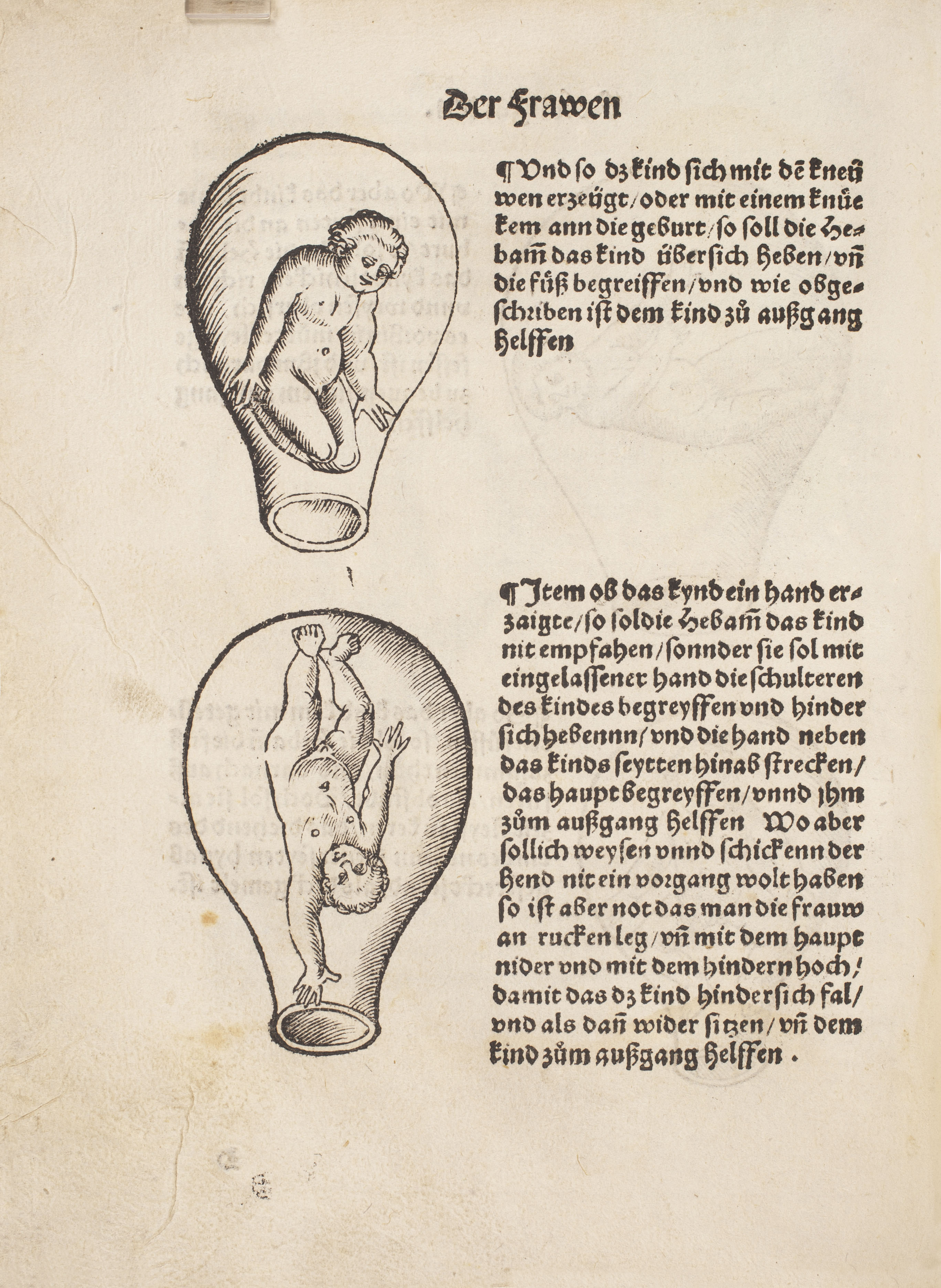
‘Der schwanngeren frawen und hebammen rosegarten’ (published 1528) by Eucharius Rösslin (?–1526)
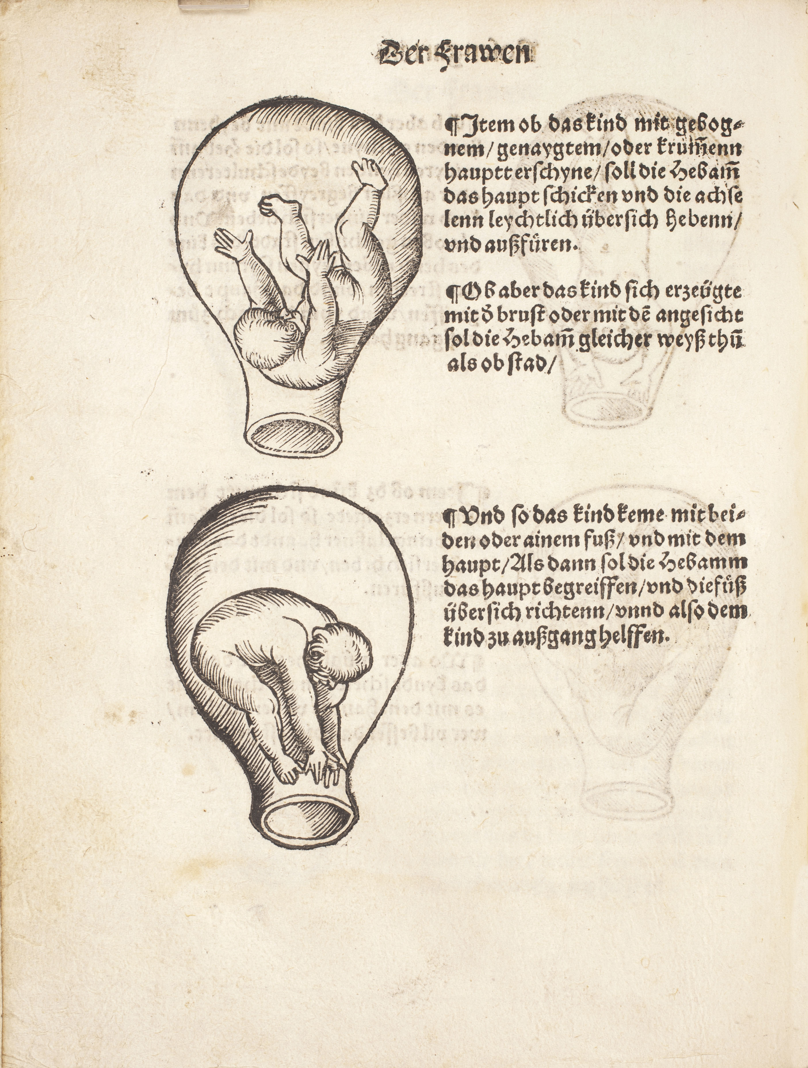
‘Der schwanngeren frawen und hebammen rosegarten’ (published 1528) by Eucharius Rösslin (?–1526)
As some of the first published images in an illustrated manual for midwives, the images of the fetus in utero in this German-language text became iconic in early sixteenth-century Europe owing to its widespread distribution in many languages and editions. In English, it appeared as The Rose Garden. Although today these images appear whimsical, we should not mistake their cartoon-like quality for the very real purpose they served practising midwives as a manual of childbirth and the many positions in which a fetus—or fetuses, in the case of twins (bottom right)—might present themselves. The presentation of various birthing positions was tremendously popular in this first generation of midwifery manuals, indicating a desire on behalf of practitioners to catalog and taxonomize fetal malpresentation.
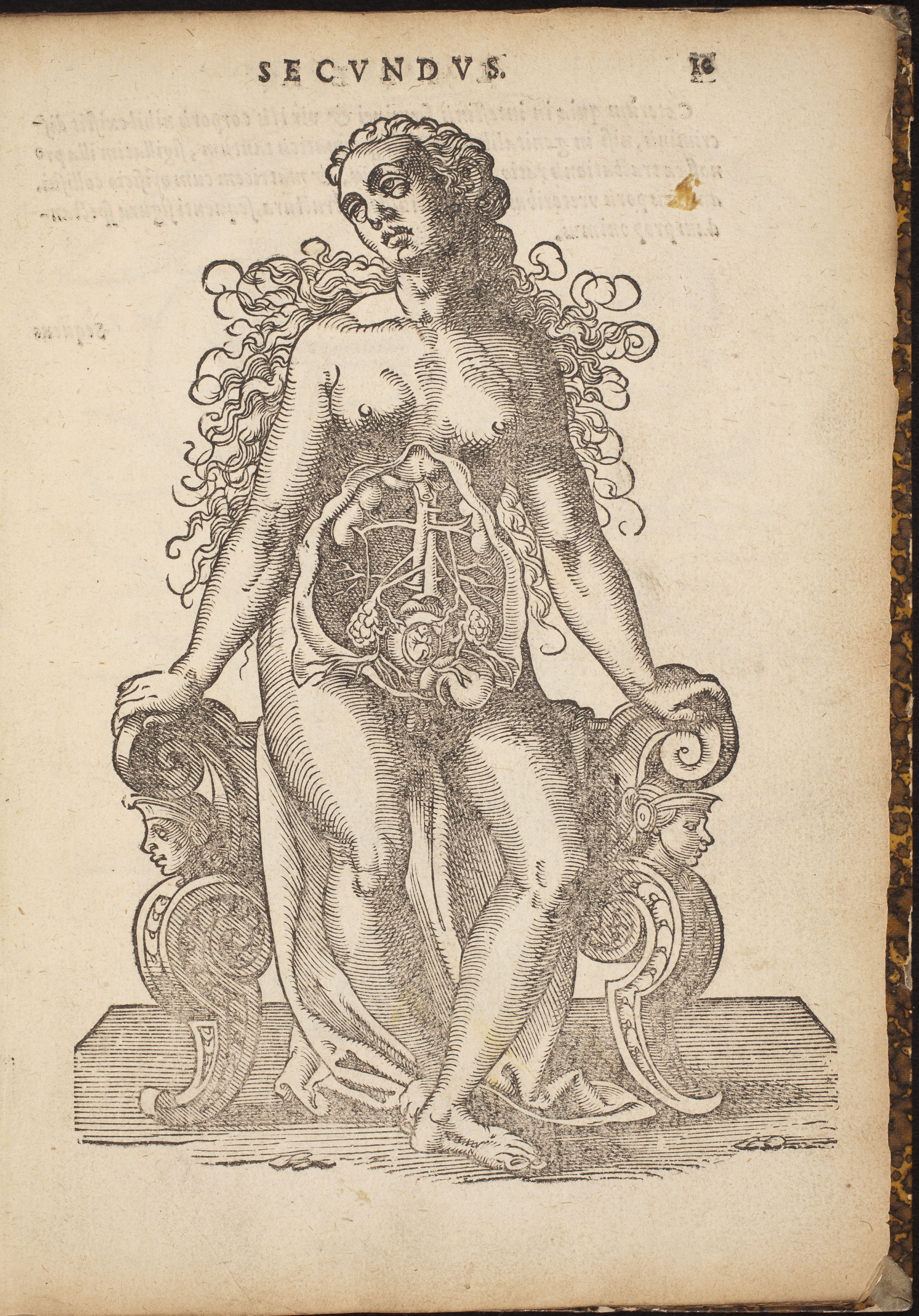
‘De conceptu, et generatione hominis’ (published 1587) by Jakob Rüff (1500–1558)
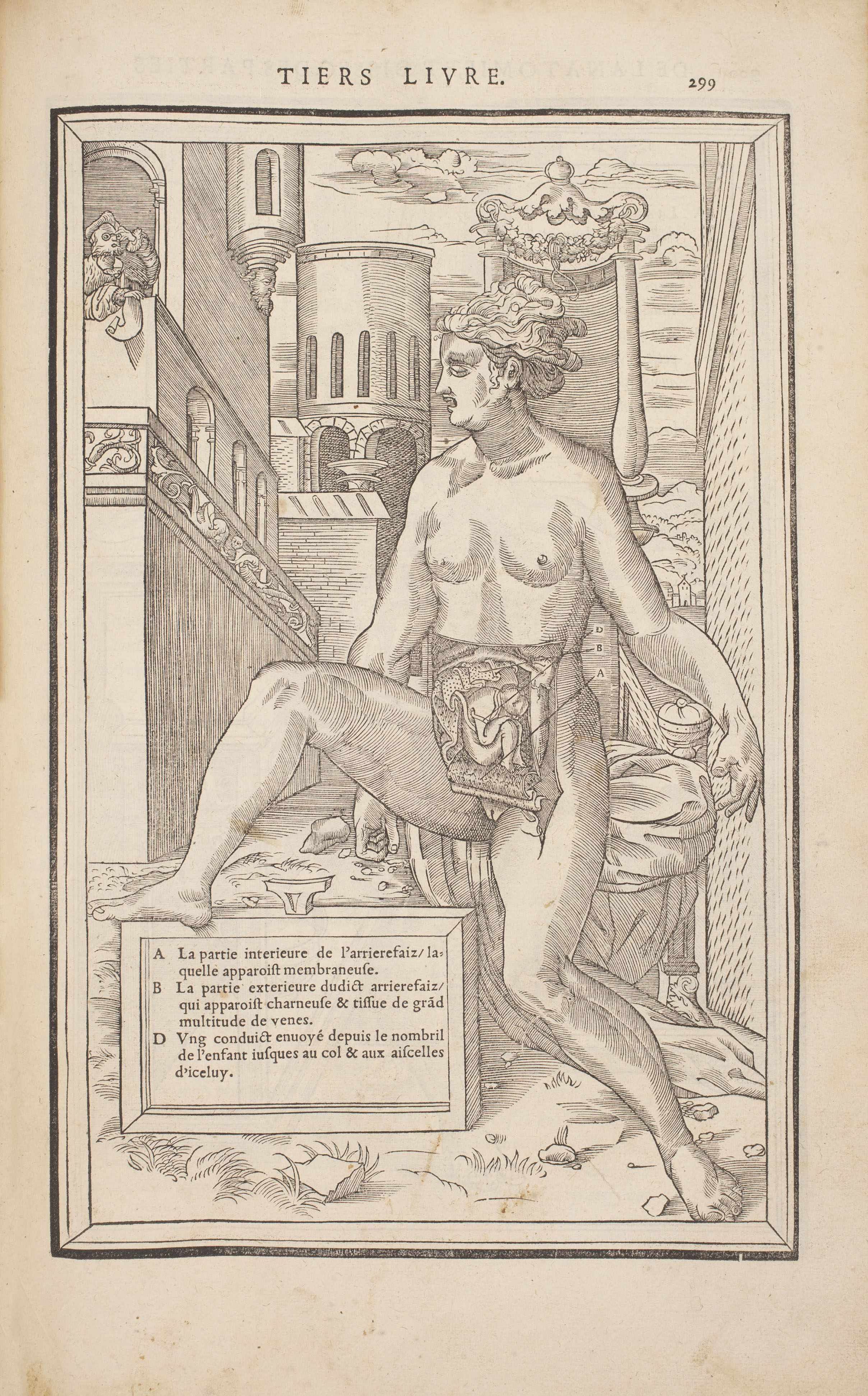
This engraving is a companion to the neighboring snapshot from another work by the Swiss surgeon, Jakob Rüff. This image of a fully embodied mother with a small fetus in utero appears in his treatise on human generation. The mother’s statue-like pose is both contemplative and resigned. The slightly wild appearance of her upper torso—a sideward gaze, exposed breasts, and flowing hair—is offset by the solidity of her lower body, with both her buttocks and arms planted on decorative furniture featuring legs with busts of two men looking outwards. In another engraving from a contemporary work, a mother might assume such a posture while delivering an infant on a stool while flanked by midwives who are assisting in the delivery. The drape on which this mother sits does not play the part of modesty cover, as in most other iconography of pregnant woman.
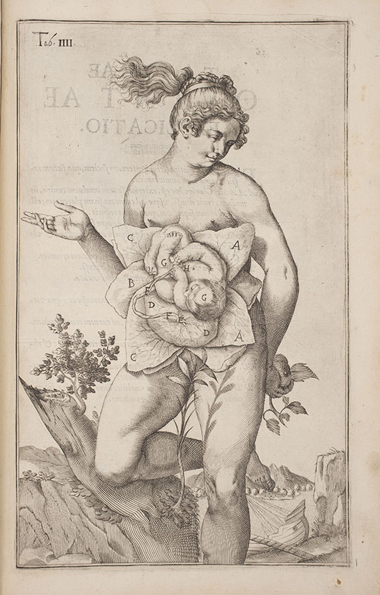
‘De formato foetu liber singularis aeneis figuris exornatus’ (published 1626) by Adriaan van de Spiegel (1578–1625)
This engraving presents a ‘blooming flower’ fetus cradled by several petals, as if to acknowledge the role of mother nature in nurturing organic life. The thin, vein-like lines on the leaves enveloping the fetus point to knowledge of fetal-maternal blood circulation and the life-giving properties of the umbilical cord. This work also points to future naturalistic mythology surrounding human conception, such as the cabbage patch and stork origins stories. Unlike many depictions, the mother is present in full. She adds new flourish to the aesthetics of Renaissance statuary—wherein women were presented as both voluptuous and muscular—with her free-flowing hair, bent knee, and leafy plant covering her genitalia in order to preserve her modesty. Unlike most classical female statues whose arms are strategically placed to cover privy parts, this mother’s right arm freely points outwards, suggesting perhaps a new ethos of demystifying the fetus in utero. The nondescript naturalistic setting was a common feature of medical atlases of this period that represented a nod to the classical world that Renaissance anatomy strived to update.
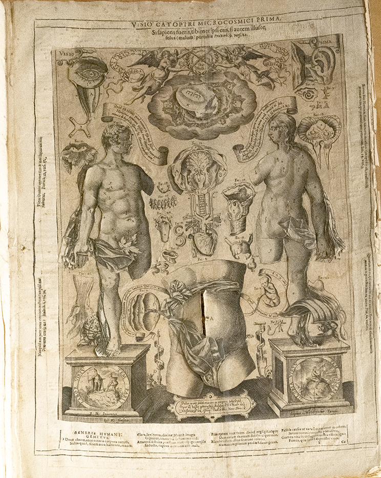
‘Catoptrum microcosmicum’ (published 1660) by Johann Remmelin (1583–1632)
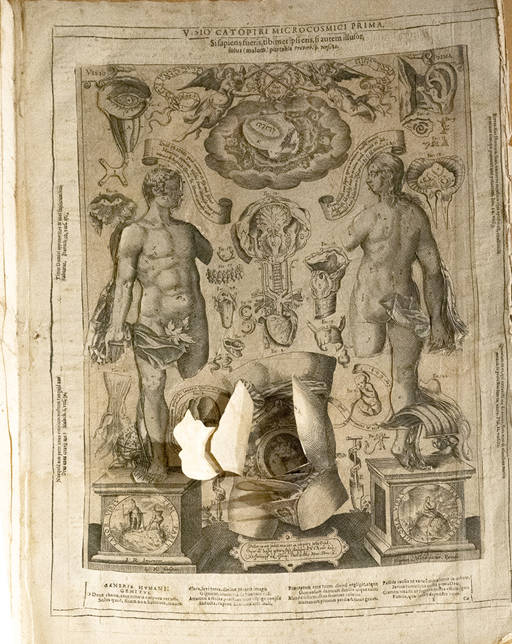
‘Catoptrum microcosmicum’ (published 1660) by Johann Remmelin (1583–1632)
This delicate and highly prized large scale “flap anatomy” provides the reader with the unique tactile experience of opening up the compartments of the male and female body respectively, as if dissecting a corpse. Remmelin’s depiction of the disembodied female abdomen pictured here (bottom center) allows students to peel back flaps in order to arrive at the near-term fetus in utero. In other editions of this work, a diabolical figure appears directly below the gravid belly, as if to signal the proximity of evil in god’s handiwork. As the title suggests, by opening up the flaps, the reader is holding a unique “mirror” onto a “small world” of the human interior. This “mirror” reflects back the mysteries and wonder of the creator. The notion of “mirror” remains a relevant motif in our secular medical world, in such apparatus as the vaginal speculum, a device composed of metal tongs designed to open the vagina for enhanced visual inspection.
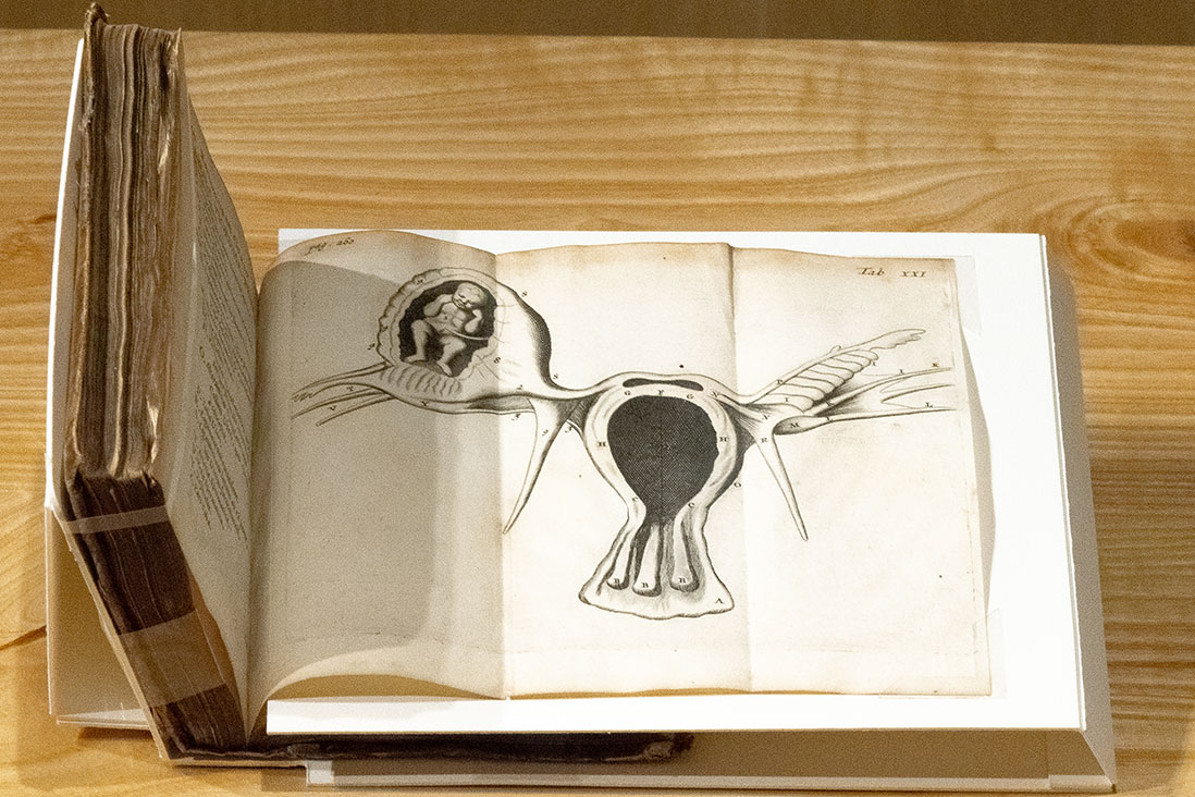
‘Regneri de Graaf medici delphensis partium genitalium defensio’ (published 1673) by Reinier de Graaf (1641–1673)
This fold-out image depicts an ectopic pregnancy in which the embryo is developing outside of the central cavity of the uterus. Here, the Dutch anatomist Reinier de Graaf provides insight into how ‘abnormalities’ in human development were both categorized and graphically presented. For most early modern anatomists, the study of abnormalities was the contemplation of marvels and prodigies. Their existence was attributed to various causes—from god’s curse to the unseemly maternal imagination. The latter was the belief that a mother’s untoward interactions with the outside world, such as witnessing the execution of criminal, might result in negative physical or psychological imprints on the fetus. By the eighteenth century, such abnormalities were generally classified as “counter-natural” occurrences that were nonetheless part of the natural world, and so, equally worthy of rational scientific study. Towards the end of the eighteenth century, abnormalities were labelled pathologies, and by the nineteenth century, the emerging field of “teratology” meant that such oddities of nature began to take on the more modern meaning of congenital abnormality or birth defect.
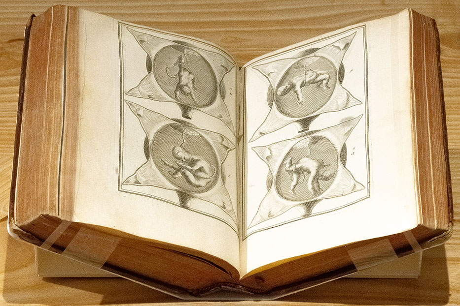
‘La pratique des acouchemens’ (published 1694) by Philippe Peu (1623–1707)
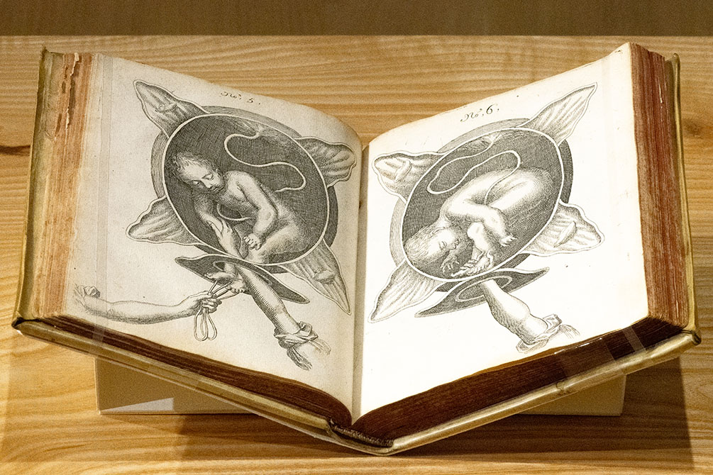
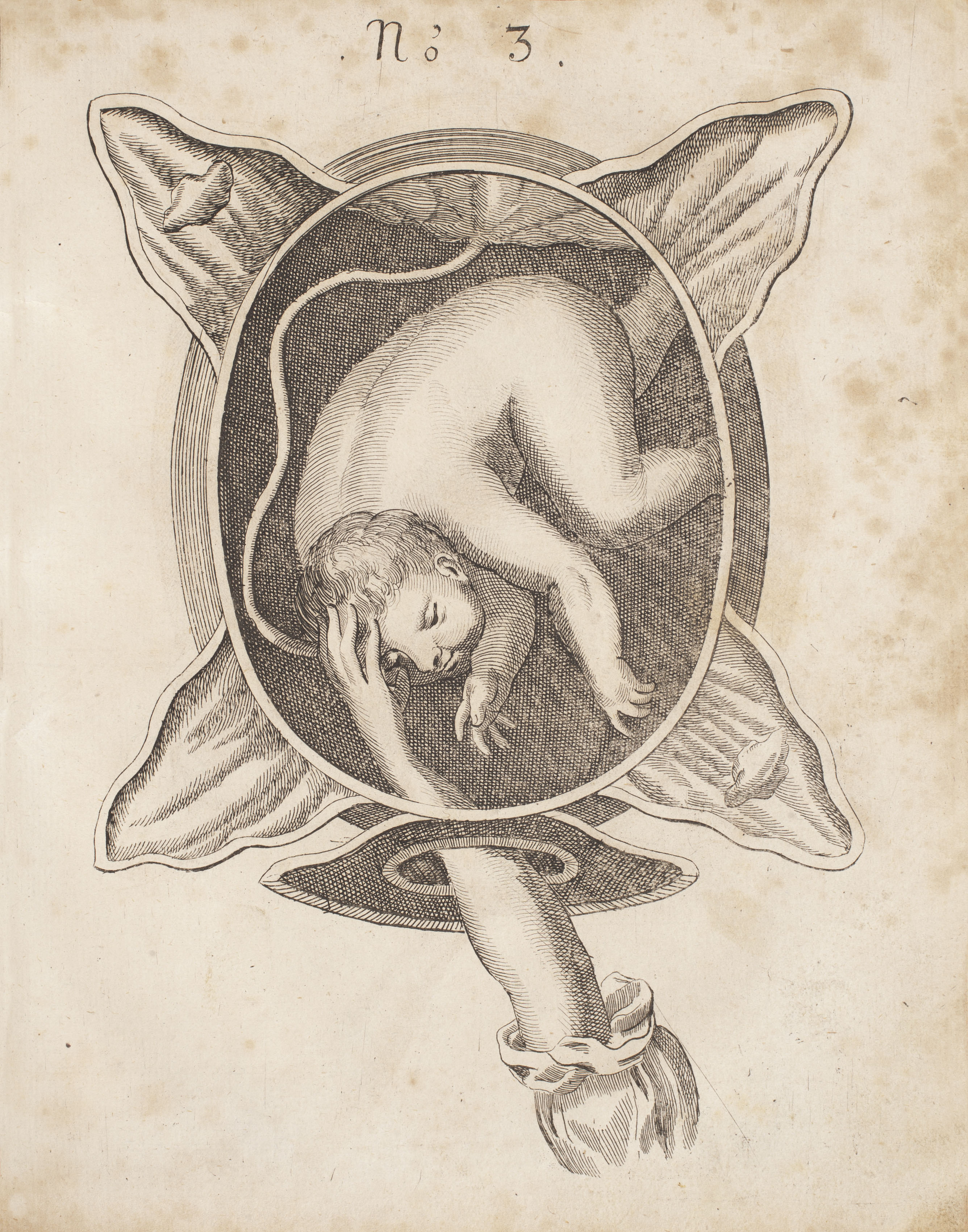
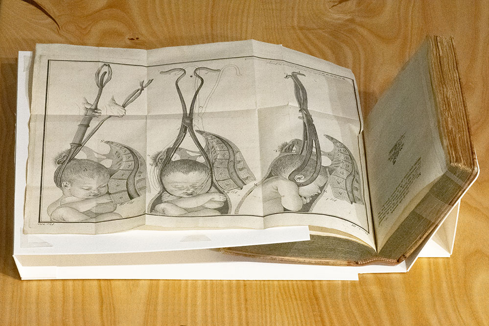
This playful series of four images from Philippe Peu’s The Practice of Childbirth depicts the fetus in seeming freefall in what would today be considered a comically spacious womb. The uterus that has been flayed open suggests that an anatomical dissection occurred, providing a rare window into the living world of the fetus. This also reveals the tension between the desire to visualize the living body in labor with the realities of gaining such knowledge through posthumous dissection of pregnant women. Herein Peu highlights several of the most common fetal positions and potential entanglements with the umbilical cord, based on his observation of several thousands births as an “accoucheur” (man-midwife) at the Parisian Hotel-Dieu birthing hospital.
; Title of work: Tractatus quatuor; Publishing information: S.l: S.n, n.d.; Call number with collection: fQM601.F2 Rare; Barcode: 108072523;")
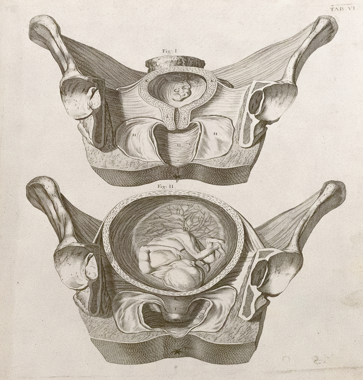
‘Tractatus quatuor’ (published n.d.) Fabricius ab Aquapendente (1537–1619)
This engraving depicts two different images of the fetus in utero: while the top (VI) shows the fetus in the uterus attached by the umbilical cord, the bottom right image (VII) reveals a near-term fetus positioned to descend the birthing canal. The Paduan surgeon-anatomist Hieronymous Fabricius (ab Aquapendente) studied under Gabriel Fallopius (1523–62), the namesake for the fallopian tubes. Like his mentor, Fabricius was a pioneer in the study of fetal development and animal-human comparative embryology.
The Art and Science of (Man-) Midwifery (1650-1750)
The Art and Science of (Man-)Midwifery
(1650–1750)
During the eighteenth-century Enlightenment, male surgeons and physicians took an increasing interest in a domain traditionally considered to be the vocation of midwives. The reasons for this interest were numerous, with some regional variation. In France, for example, state pressures to reduce infant mortality resulted in King Louis XV (1710–74) providing Mme du Coudray (1712?–94) with a royal pension in order to tour the country and improve birthing practices through her innovative teaching methods. She trained male and female students in various birthing postures on a simulative mock-woman made of real pelvic bone and textiles that she referred to as both her “phantom” and “machine.”
Mme du Coudray worked alongside, and sometimes in opposition to, a number of male practitioners who were making professional claims to childbirth. They did so primarily, though not exclusively, by way of instruments, which they argued were superior to human hands in delivering infants in the case of fetal malpresentation. French practitioners, like François Mauriceau (1637–1709) and Jean-Louis Baudelocque (1745–1810), pioneered new instruments and identified professionally with their surgical ingenuity. Extractive devices like forceps were commonly used to assist in delivery, while in more radically obstructive cases, such ominous-looking devices as the “crochet” (a sharp metal hook) or the “tire-tête” (a corkscrew-like implement) were implemented. While these tools often saved the mother, they sacrificed the fetus during removal. These instruments and their use increasingly appear in illustrated midwifery manuals, further medicalizing the fetus in utero.
The intrusion of male medical authority in midwifery extended across Europe and in both America and colonies in the Americas. The male takeover of traditional midwifery was not without opposition, however. The English midwife Elizabeth Nihell (1723–76), who had trained at the prestigious Hôtel-Dieu of Paris maternity ward, was a staunch adversary of male “instrumentarians.” She complained that they applied excessive, violent force in childbirth through their cold metal tools in cases that simply required patience and manual dexterity. Other women took male encroachment on their profession as motivation to improve and expand their own techniques and textual output.
The German midwife Justina Siegmund (1636–1705) stands out as an enterprising practitioner who overcame the jealousy of male colleagues while also presenting novel solutions to complicated birthing positions in her illustrated manual Court Midwife (1690). In this work, Siegmund presents an ingenious two-handed technique using a sling, in order to rotate the fetus in utero presenting its shoulder into the more optimal head-first position. This method demonstrated that instrumental intervention and hand-based delivery were not inherently oppositional, and moreover, that textile-based instruments like slings also had their place in infant delivery alongside rigid metal ones. Her inclusion of the latest embryological and anatomical engravings from experts in the field, including the Dutch physician Govard Bidloo (1649–1713) featured elsewhere in this exhibition, also showed that Siegmund was at the cutting edge of her field.
The Art & Science of (Man-)Midwifery (1650–1750)

‘Des maladies des femmes grosses et accouchées’ (published 1668) by François Mauriceau
‘Die chur-brandenburgische hoff-wehe-mutter’ (published 1690) by Justina Siegemund (1636–1705)
As the first female midwife to publish a midwifery manual, The Court Midwife in 1690, Justina Siegemund depicts a variety of birthing scenes, which instruct thoroughly without appearing immodest or too invasive. In these engravings of a breech birth, the disembodied arms of the midwife reach towards an angelic baby. The baby is serene, plump, and with enough room to move, while the uterus of the mother is completely severed from its anatomical background. The precision of the feminized hand, as well as the instrumental string, nonetheless provide enough detail to make this a useful teaching tool for future midwives. Caption written by Antonia Willnow
‘The art of midwifery improv’d’ (published 1716) by Hendrik van Deventer (1651–1724)
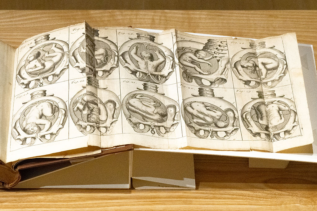
In this English-language translation of the Dutch man-midwife Hendrik van Deventer, we see a fold-out showing ten different fetal positions, including both “natural” (head-first) and “counter-natural” breech (feet-first) ones. Although the near-term fetus has room to maneuver in utero, the spaciousness of the uterus relative to the fetus has been reduced when compared to works from the mid-sixteenth-century Renaissance. This suggests that dissections of gravid uteruses have sharpened practitioner’s notions of the geometry of the uterus, as well as pelvis, and fuelled their goal of rationalizing childbirth through more refined understandings of pelvic capacity in delivery. The sheer number of fetal eventualities portrayed confirms this taxonomic goal. The book’s title suggests that midwifery was a field ripe for improvement—at least, according to the generation of male practitioners who sought to introduce their surgical tools and anatomical knowledge to the domain of the midwife.
‘Observations importantes sur le manuel des accouchemens’ (published 1734) by Hendrik van Deventer (1651–1724)
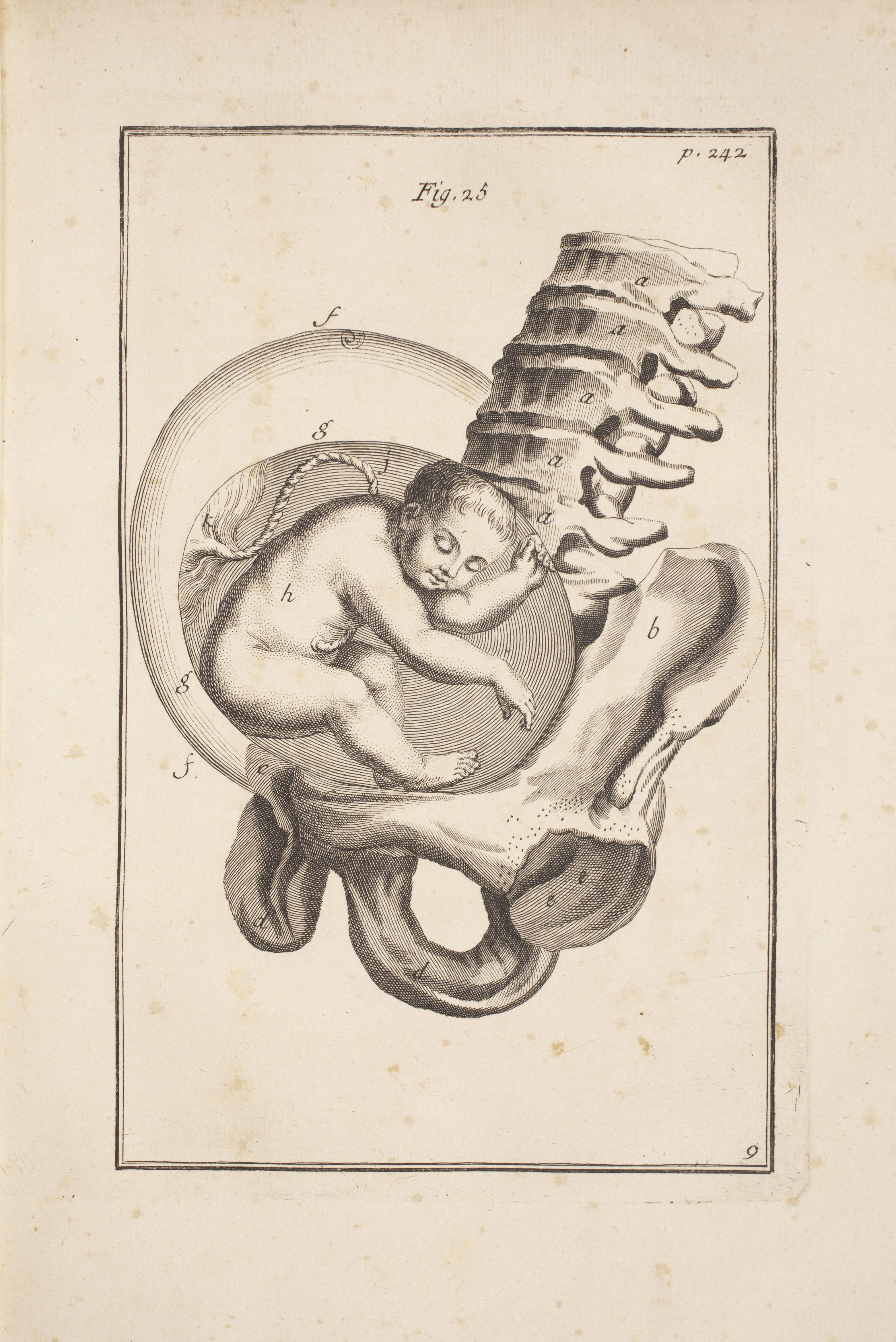
This image provides a fitting companion to Hendrik van Deventer’s neighboring fold-out presenting multiple fetal positions. Here, the lone fetus is depicted in the uterus of an all-but-disembodied mother, with emphasis placed on how her osteological features—including the positions of vertebrae and pelvic bones—interact with the fetus in ways that both facilitate and sometimes obstruct childbirth.
‘Die königl’ (published 1741) by Justina Siegemund
‘Die königl’ (published 1741) by Justina Siegemund
Figures 2 and 3 show a midwife’s arm adjusting the position of the fetal head to allow for an easier birth. Her arm is cropped right under the elbow, the sleeves she wears are billowing and graceful, as if she were doing God’s work, and the gentle touch on the baby’s head mimics that found in Michelangelo’s The Creation of Adam. The uterus is anatomically isolated—there is no placenta or additional tissue, and the mother is otherwise absent. This reveals a distaste for the female body, while heightening the importance and religious significance of birth. Caption written by Antonia Willnow
‘Observations sur la pratique des accouchemens, naturels, contre nature, & monstrueux’ (published 1748) by Cosme Viardel
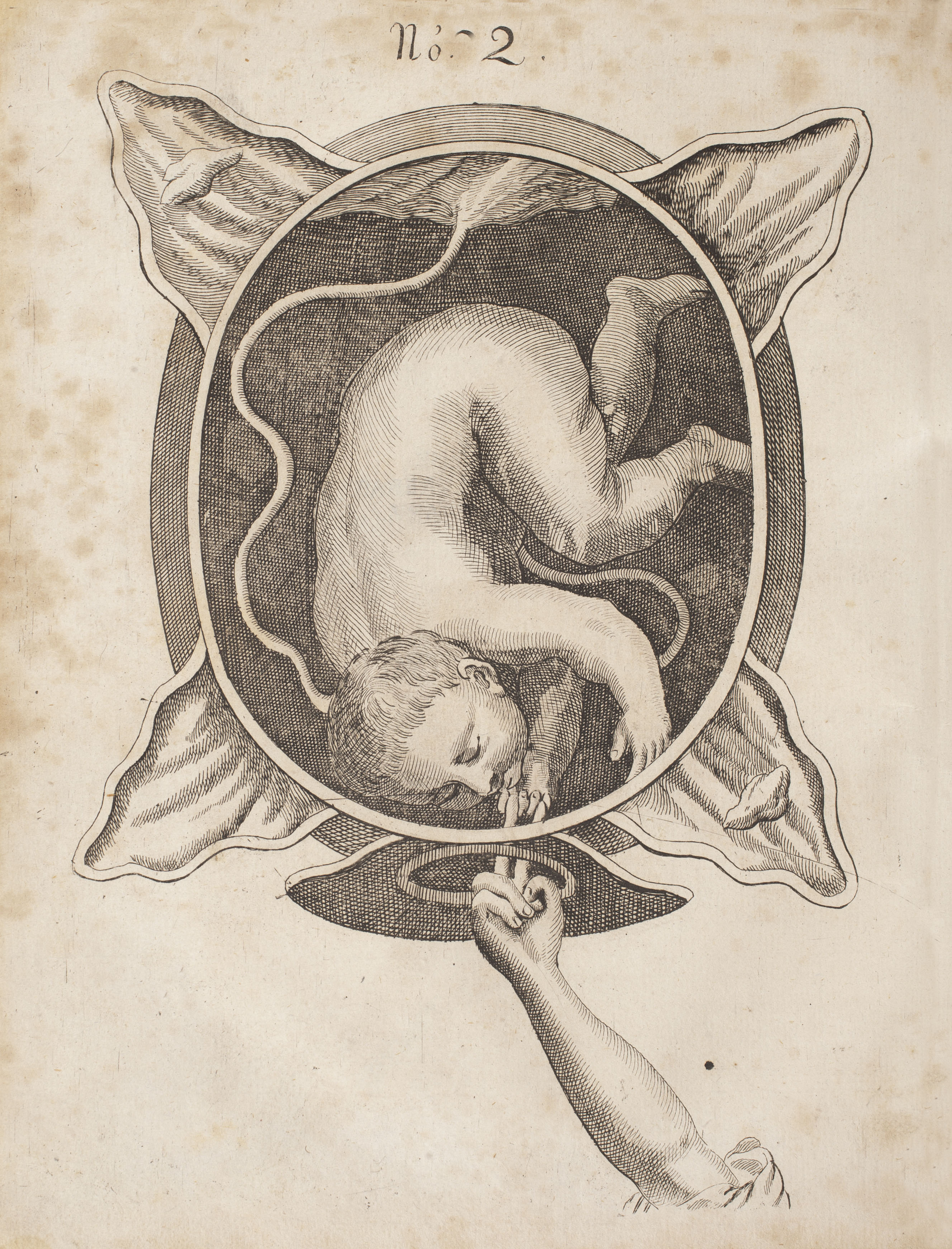
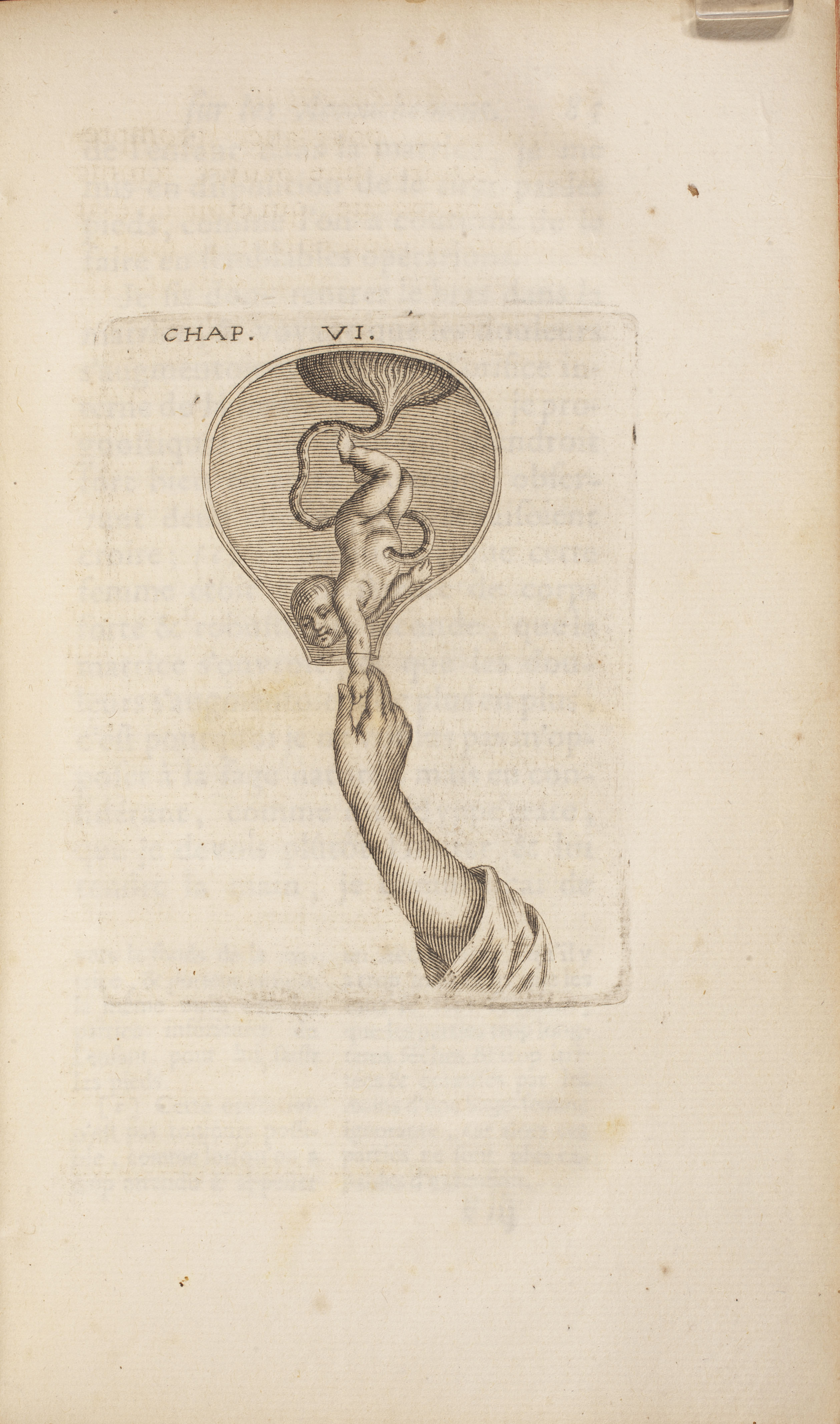
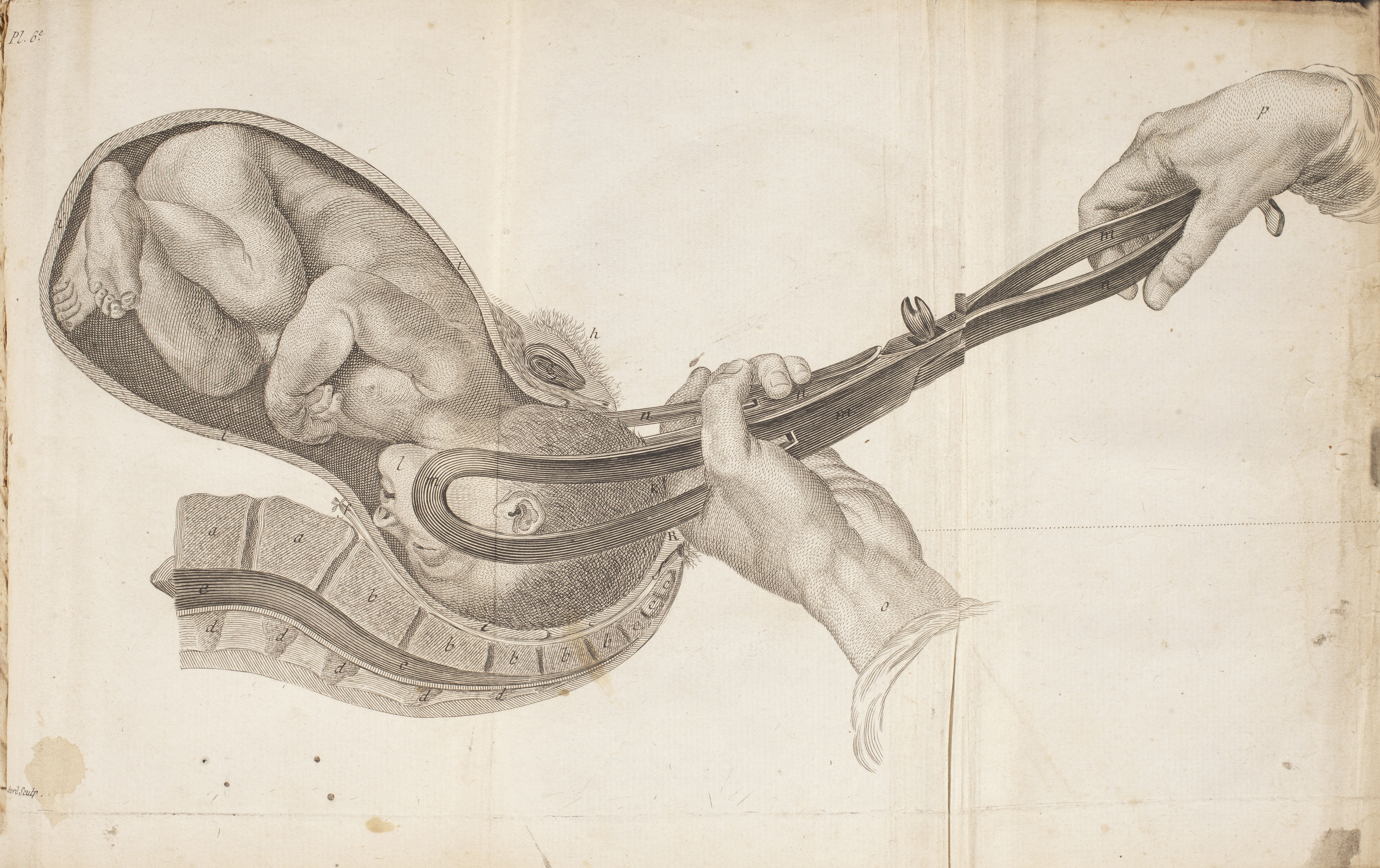
This engraving presents a full-term fetus in a roomy womb at the moment of birth, with the helping and larger-than-life hand of a presumably male practitioner guiding the fetus to life ex utero. The text accompanying the image explains that situations in which the infant presents its arm first are among the most difficult deliveries, given they do not permit recourse to any variety of “operation.” In this context, an operation was understood to mean instrumental intervention. It thus falls to the dexterous, experienced practitioner to reposition the fetus into the ideal head-first birthing position.
‘An essay towards a complete new system of midwifery’ (published 1751) by John Burton
‘Instructions succintes sur les accouchemens en faveur des sages-femmes’ (published 1770) by Joseph Raulin (1708–1784)
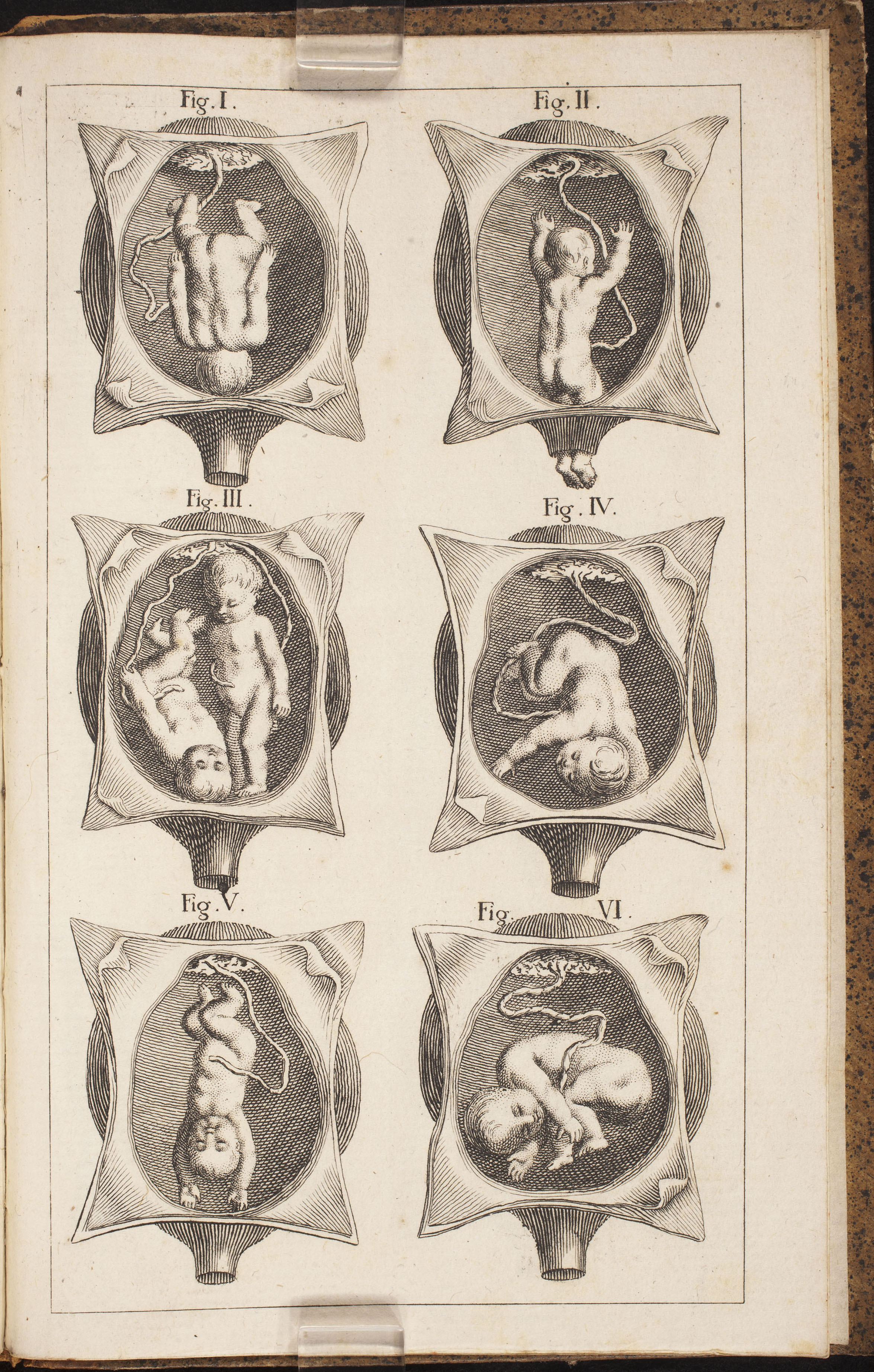
This small format work by physician to the French King Louis XV (1710–74) presents six different fetal positions in utero, including a set of twins facing opposite directions. The spaciousness of the uterus presented here is in contrast to the naturalistic works of contemporaries like William Hunter (1718–83) whose Anatomy of the Gravid Human Uterus presents more realistic full-term fetuses in utero with no room to maneuver.
‘L’art des accouchemens’ (published 1781) by Jean Louis Baudelocque (1745–1810)
If the Scottish man-midwife William Smellie (1697–1763) has gone down in history as the “father of British midwifery,” then Jean-Louis Baudelocque earns the French equivalent title. He was a keen instrument maker who sought to rationalize childbirth by introducing new tools for delivery. His external “pelvimeter” was a predictive anthropometric device designed to measure a mother’s pelvic capacity, in order to determine if other instruments were required in a difficult delivery. In this striking fold-out image, we see a male practitioner’s hands delivering a naturalistic-looking full term infant using curved forceps, a prototype introduced by Baudelocque to facilitate extraction. These features—the realistic fetus, the reduction of the mother to the amniotic sac and a spinal column interacting geometrically with the fetal skull, and the recourse to instruments even in the ideal head-first birthing posture—reveal the primary aims of man-midwifery, to medicalize childbirth from a surgical and interventionist perspective. What this image does not portray is equally telling of these aims. The virtual erasure of the mother, and especially the absence of the birth canal—which is the only conduit for the forceps—tells us about her silencing in the new regime of childbirth. As surrounding sources attest, the disembodied mother was normalized in fetal iconography in this period.
‘Tabulae anatomicae ex archetypis egregii pictoris’ (published 1788) by da Cortona Pietro (1596–1669)
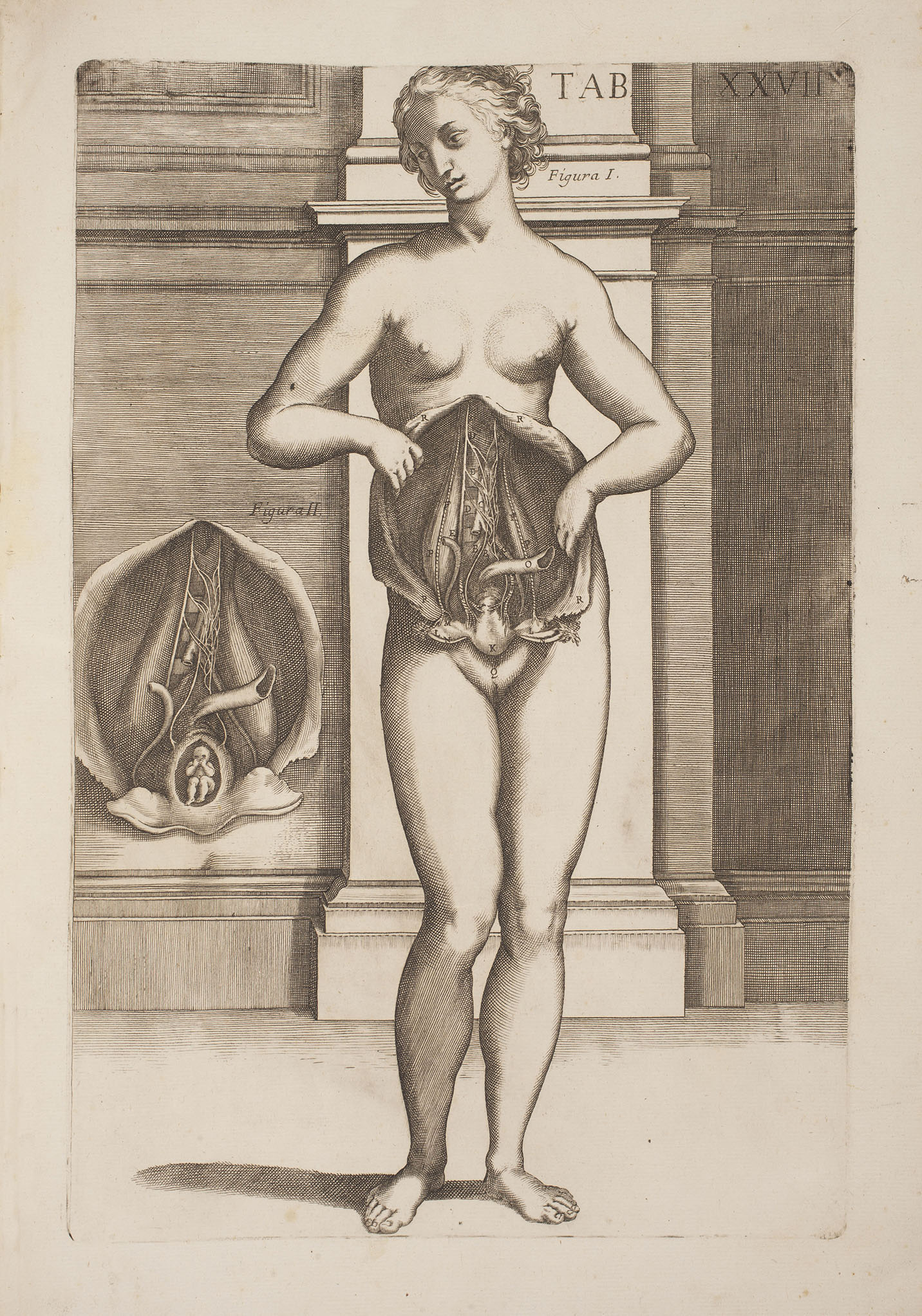
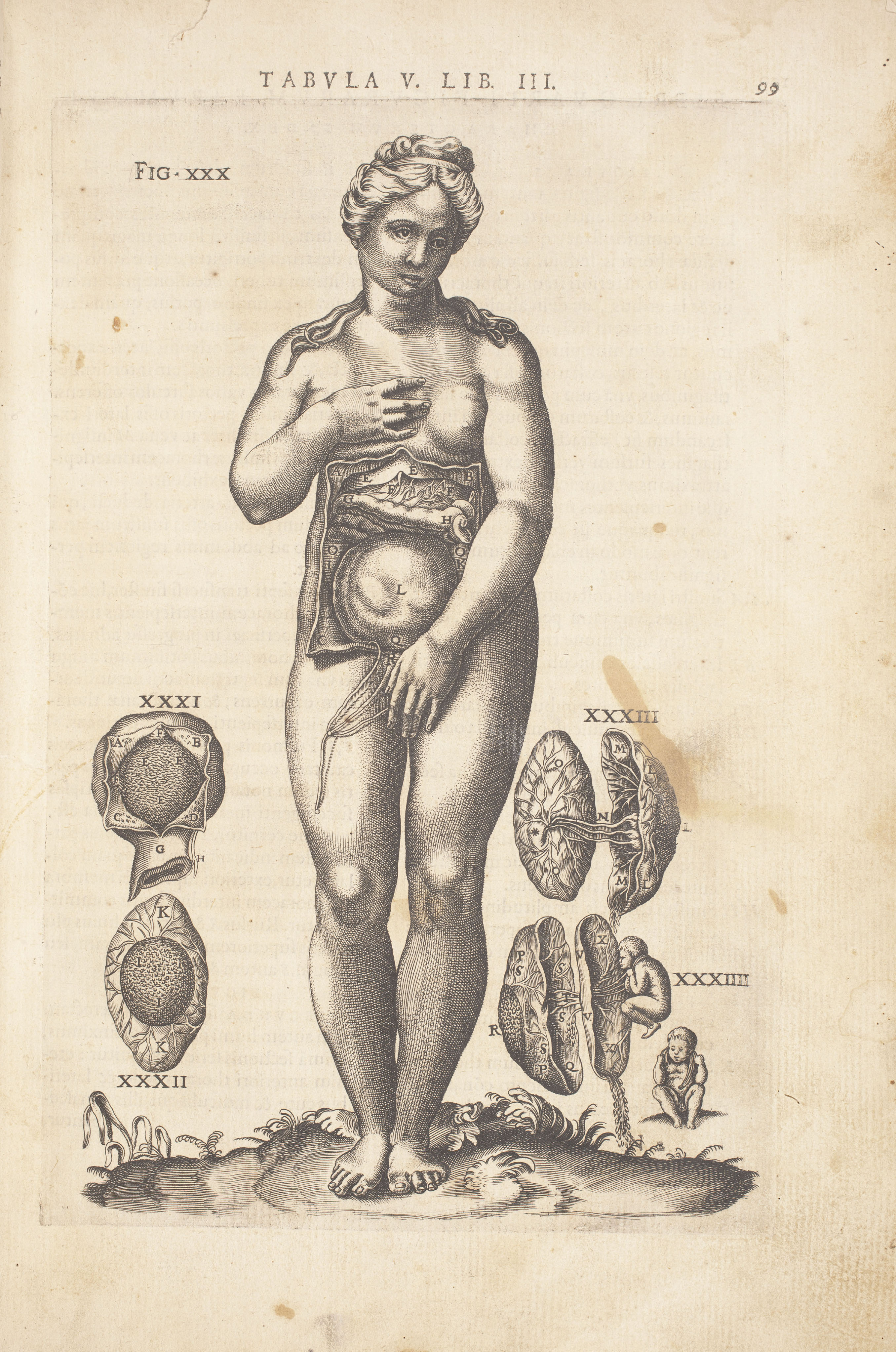
In this anatomical table, we see a lifesize and lifelike woman undertaking the impossible task of flaying her own abdomen, in order to unveil her viscera and inner organs of generation. To the left, we see a close-up of the open abdomen in which rests a tiny fetus-containing uterus. The maternal posture is similar to those featured in other anatomical works dedicated to the human body, most of which approximate Renaissance female statues, which paid homage to classical statuary and were often referred to as Venuses. While this is a 1788 edition of the work, its iconographic style dates its original publication to the mid-seventeenth century.
From Midwifery to Obstetrics (1700-1900)
From Midwifery to Obstetrics
(1700–1900)
By the early nineteenth century, the man-midwife had asserted his professional authority in a field that was transforming into a science known as obstetrics. The man-midwife, or “accoucheur” as he was known in France, rose to prominence over the female midwife, in a process that transformed the profession and medicalized childbirth.
Although one might expect in this period of disciplinary upheaval, the continued exclusion of the mother or her disembodiment in graphic anatomy, nineteenth-century visual depictions often present a re-embodied mother. This was a departure from the Enlightenment period, when “butcher shop meat” was a common mode of maternal portrayal. Presenting an embodied mother provided an opportunity to convey the Victorian virtues of gentility and modesty. However, if the mother now appeared more fully and transparently, she was nonetheless presented with moral and social objectives in mind.
In this regard, Enlightenment and Victorian visuals shared in a prescriptive discourse of pregnancy in which the female body should be presented through either the male medical gaze or a broader societal gaze. In the case of the Enlightenment practitioners focused on ‘rationalizing’ the body, the mother was disembodied from the process in order to assert their authority or to do the ‘social good’ of improving childbirth and decreasing infant mortality. Once this process was near completion and the ‘science of childbirth’ routinized in the nineteenth-century hospital environment, attention arguably turned to the re-embodied mother, in order to reflect the social pressure to safeguard reproduction as a means to ensure a workforce during the Industrial Revolution.
The move towards a hierarchical and male-dominated vision of childbirth, and the extension of graphic representations of the fetus in utero to the moral and social realms, was not to the exclusion of female practitioners, however. French women like Mme du Coudray (c. 1712–94) and Mme Boivin (1773–1841), for instance, found themselves at the cutting edge of disciplinary change. Unusually for a woman in this period, Mme du Coudray obtained a pension from King Louis XV (1710–74) in order to undertake a twenty-five-year mission to educate both men and women in the art of birthing. Mme Boivin wrote textbooks, including her multi-edition Memorial on the Art of Childbirth (1817), trained cohorts of students at the Paris Maternity hospital, and invented instruments like an internal “pelvimeter,” a diagnostic tool designed to predict difficult labors due to a too-narrow pelvic passageway.
Finally, depictions of the fetus in utero in this period of disciplinary ferment fed off changes in imaging technology. Copperplate engraving, which had replaced woodcut printing, persisted in this period. It is a technique by which an image is drawn by an illustrator before being etched onto a copperplate by an engraver, and then inked for mechanical reproduction. Lithograph techniques were also used, which permitted printing from either a stone or metal plate using chemical processes. Interactive flap anatomies and life-size posters designed for classroom use also contributed to visualizing the fetus in utero. The traditions of artisanal drawing, etching, and engraving would all be revolutionized by the end of this period, with the advent of photography and the x-ray.
From Midwifery to Obstetrics (1700–1900)
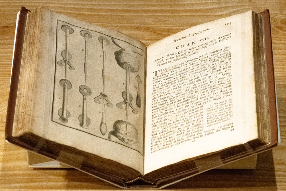
‘The diseases of women with child’ (published 1710) by François Mauriceau (1637–1709)
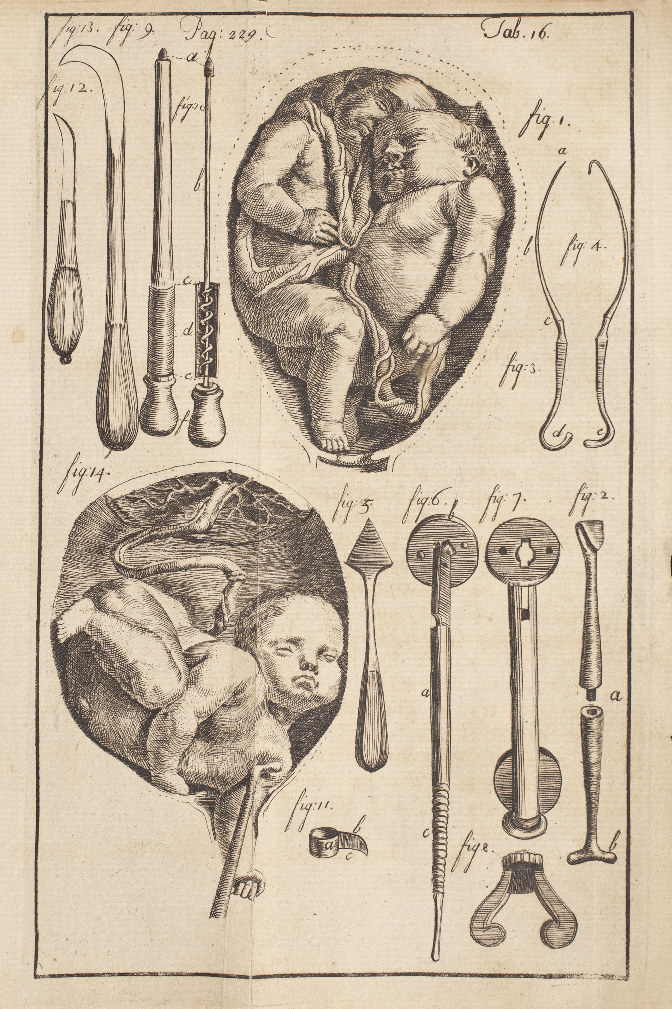
This work devoted to female gynecological diseases was originally written in French by François Mauriceau, who apprenticed in midwifery at Europe’s foremost maternity school, the Hôtel-Dieu hospital of Paris. As a surgeon rather than the loftier physician, he often surprised in his anatomical knowledge of the fetus, gravid uterus, and female pelvis. Mauriceau invented a “tire-tête” instrument designed to remove a child who has either perished in utero or is near death. He argues in the text accompanying this engraving that, when the mother’s life is at risk, the practitioner “need then make no scruple” in using such instruments to extract the fetus.

‘Le guide des accoucheurs’ (published 1743) by Jacques Mesnard (1685–1746)
This “accoucheur” or man-midwife from Rouen, France published A Guide for Man-Midwives in 1743. He pioneered surgical instruments for performing the then-dangerous Caesarean operation, which he shared and illustrated in this Guide. Pictured here is a purpose-made birthing bed, one of a number of prototypes developed in this period by male practitioners to place the mother in a recumbent position. This facilitated both the male surgeon’s gaze into the birth canal and any instrumental manipulation that might be deemed necessary in delivery. The invention of the birthing bed was in breaking with past practice favored by midwives, who encouraged mothers to give birth sitting upright on purpose-made stools.
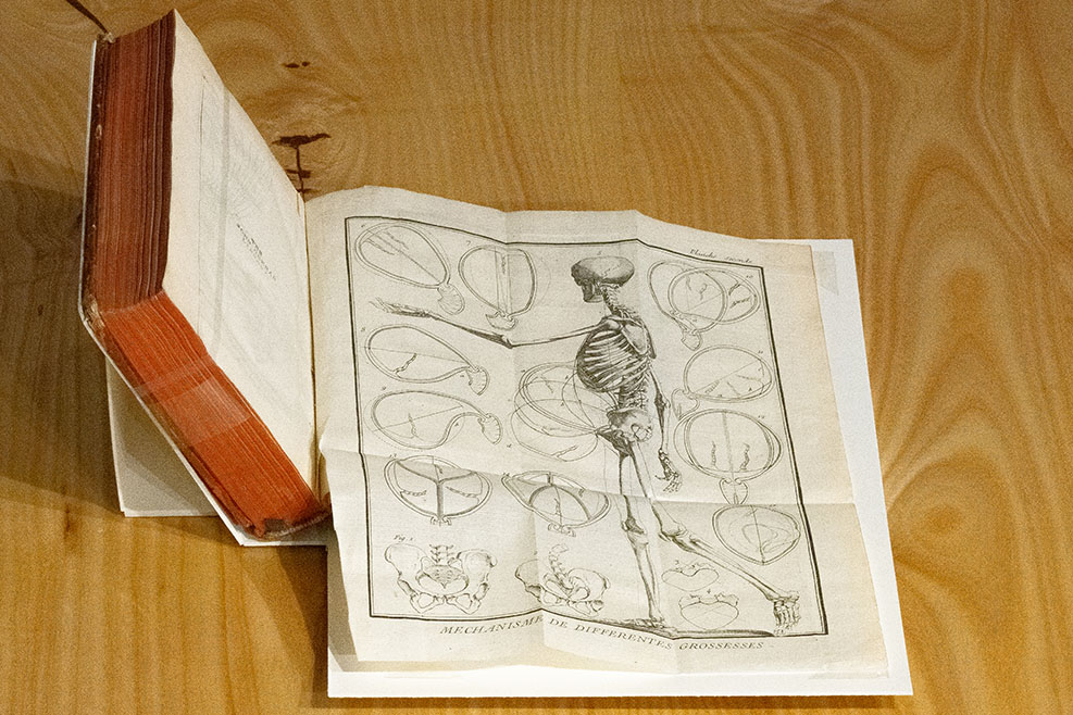
‘L’art des accouchemens’ (published 1761) by André Levret (1703–1780)
This engraving depicting a female skeleton, with silhouettes outlining how she will carry her fetus at various stages of pregnancy, is a companion to the nearby image of Levret’s curved forceps. The pelvises presented at the bottom left indicate practitioners’ interest in determining pelvic capacity prior to childbirth. The label reads “Mechanisms of different pregnancies,” suggesting that the pregnant mother is akin to a machine that can be rationalized.
‘Abrégé de l’art des accouchements’ (published 1777) by Angéglique Marguerite Le Boursier du Coudray (ca. 1714–1794)
This French guide for midwives served as a pocket-sized Guide to the Art of Midwifery. The guide provides technical as well as bedside manner instruction for midwives-in-training. Mme du Coudray’s conversational tone avoids condescension, and most surprisingly, she avoids euphemistic language. Her use of learned terminology to describe female genitalia and reproductive organs indicates her goal to stamp out the anatomical ignorance associated with country midwives. Du Coudray uses diagrams of the fetus in utero to help her reader see both the anatomical and emotional factors at play during pregnancy. The delicate floral imagery in the hand-colored paintings serves to illustrate the fragility and vulnerability of pregnancy, both for mother and fetus. Du Coudray used copies of this guide during her career training midwives across Europe, which typically began with hands-on manipulation of her mechanical birthing dolls. Next, this guide served as a transitional tool towards learning how to provide emotional and physical support to patients. Implicit in du Coudray’s emphasis on the technical alongside the soft skills of her craft lies the argument that midwifery is both an art and a science. Caption written by Elizabeth Crowdus
‘Observations sur les causes et les accidens de plusieurs accouchemens laborieux’ (published 1780) by André Levret (1703–1780)
Like many of the man-midwives who emerged in the eighteenth century, André Levret was French-born and -trained. He was the rough contemporary of the famed Scottish man-midwife, William Smellie (1697–1763), and was considered a pioneer alongside Jean-Louis Baudelocque (1745–1810) in the mechanization of childbirth that was in vogue in this period. He specialized in counter-natural or difficult births, such as the breech position, and is credited with adding a curvature to birthing forceps—as pictured here—to facilitate infant delivery.
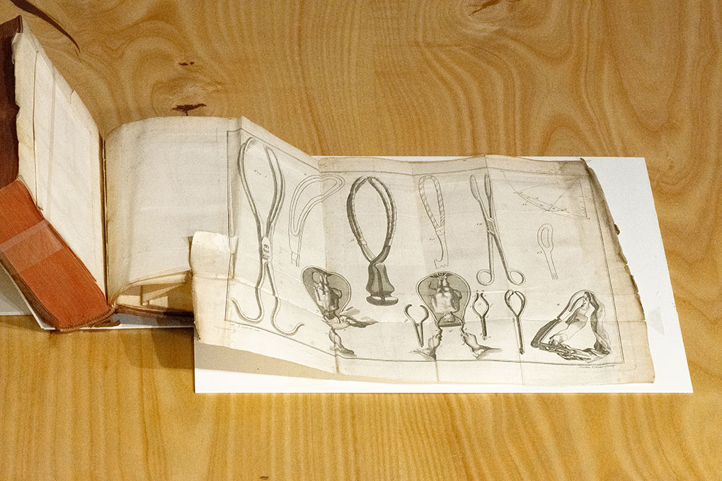
‘Traité sur divers accouchemens laborieux’ (published 1782) by George Herbiniaux (1735?–1811?)
This fold-out engraving appeared in the military surgeon and man-midwife George Herbiniaux’s Treatise on Diverse Difficult Births. He advocated use of the “levier” or lever, an instrument more familiar in the Flemish Netherlands than in France, where he practiced—and where the more intrusive forceps had gained widespread acceptance. Both of these instruments were used in cases of difficult births that were known in this period as “counter-natural.”
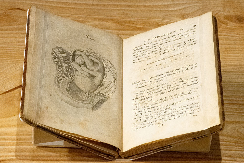
‘A set of anatomical tables’ (published 1793) by William Smellie (1697–1763)
This pocket book engraving presents a full-term fetus positioned in the uterus “when labour is somewhat advanced.” The different sets of dotted lines outlining the fetus indicate the thickness and position of the uterus both before and after the “waters” have broken. Such features, when combined with the severed thigh of the mother and her virtual disembodiment, indicates Smellie’s fetus-oriented approach to childbirth and his attempt to rationalize delivery.
‘A compendious system of midwifery’ (published 1853) by William P. (Potts) Dewees
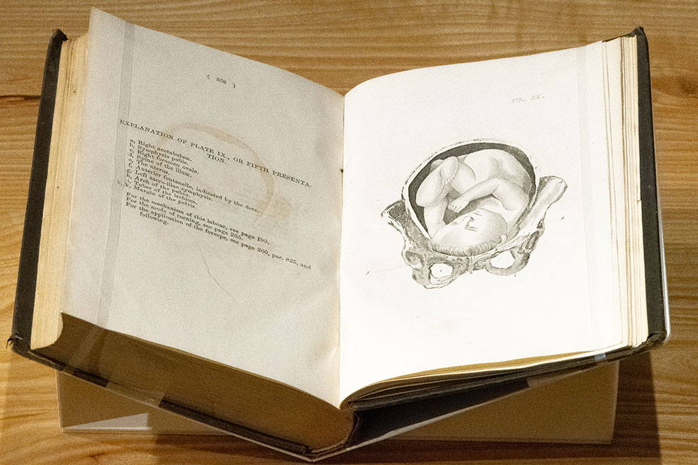
This engraving appearing in the multi-edition midwifery textbook by the Philadelphian obstetrician William Potts Dewees presents a full-term baby ready for delivery. The labelled parts of the mother’s pelvis interacting with the fetal skull indicate that osteological anatomy was of primary concern for delivery, while the accompanying text indicates that “this presentation is not attended with more difficulty” than similar head-first births. It was said of Dewees “that no parturient woman of the time considered herself safe in other hands,” which may be owing in part to his concern with pain management in childbirth.
The Consolidation of the Male Medical Gaze (1800–1900)
The Consolidation of the Male Medical Gaze (1800–1900)
Male-driven medicine was consolidated in the new space of the hospital, where old traditions of birthing were whitewashed, both figuratively and literally, by the sanitized clinical environment. Along with obstetrics, the emerging discipline of gynecology cemented male dominion over the female body. The result was the emergence of the male medical gaze, which asserted a new visual advantage over and control of patients in ways that reduced their agency when compared to earlier periods.
The cultivation of the nineteenth-century male medical gaze in the clinic was a continuation of the male takeover of midwifery that began during the Enlightenment. This gaze was facilitated by the introduction of new norms in the birthing chamber that allowed the practitioner to easily visualize, access, and interact with female reproductive anatomy. The moves towards reclinable birthing beds and gynecological examination tables favoring a recumbent position, often with inbuilt stirrups and arm restraints, facilitated practitioner control over an increasingly vulnerable patient. This was in contrast to the upright posture in which most women gave birth in the age of midwifery, where purpose-built stools were the norm.
The development of illustrated medical textbooks was another aspect of the consolidation of the male medical gaze in obstetrics. With several exceptions, including the French midwife Mme Boivin (1773–1841), male physicians set the iconographic agenda and directed teams of artisans accordingly. The main trace of their power position in curating the visual archive of the fetus in utero comes in the form of hands—hands that perform digital examinations, that wield instruments, that restrain patients. The French obstetrician Adolphe Pinard (1844–1934), for instance, was applauded for his dexterous use of palpation, the technique of manually ‘feeling inside’ the pregnant body from the outside in order to assess fetal position. Yet far from being a new technique, palpation is simply another way of describing the hands-based repertoire traditionally used by the midwife in diagnosis and delivery.
The obstetrical textbook is as interesting for what it visualizes as what it does not. Indeed, underlying many nineteenth-century depictions of the fetus in utero is a voyeuristic element. Interactive flap anatomies, which originated in the Renaissance, were nonetheless presented in new ways in this period: large-scale, full-color lithograph flaps invited readers to peel back numerous layers of the female interior in order to peer through the transparent “curtain” that is the amniotic sac. In this sense, the modesty veil of the early modern period approximates a Victorian era “peep show” curtain. The presence or absence of pubic hair, bare breasts, and external genitalia may also reveal a pleasure-seeking undertone attesting to the male prerogative of the new obstetrical textbook tradition.
The Consolidation of the Male Medical Gaze (1800–1900)
‘Mémorial de l’art des accouchements’ (published 1812) by Marie Anne Victoire Gillain Boivin (1773–1841)
This work by the celebrated French midwife, Mme Boivin, is arguably the first book of obstetrics, the new branch of medical science with a clinical bent that came into prominence in the early nineteenth century in new and refurbished hospital environments outfitted with maternity wards and teaching wings. In addition to practising and teaching midwifery, Boivin engaged in original anatomical research, invented obstetrical instruments like the “pelvimeter” (an internal diagnostic tool designed to ascertain pelvic capacity), and wrote such books as this, Memorial for the Art of Childbirth. Although open to instrumental intervention, the engravings pictured here clearly demonstrate Boivin’s commitment to manual dexterity in infant delivery.
‘Obstetric tables’ (published 1833) by G. (George) Spratt
‘Obstetric tables’ (published 1833) by G. (George) Spratt
The Obstetric Tables were designed as a pedagogical aid for male students. This hand-colored lithograph plate features flaps that, when lifted, give the impression of ‘dissecting’ the body through several layers that move the viewer from the outside to the interior of the pregnant female body. The final view brings the viewer to a transparent amniotic sac looking in on twins.
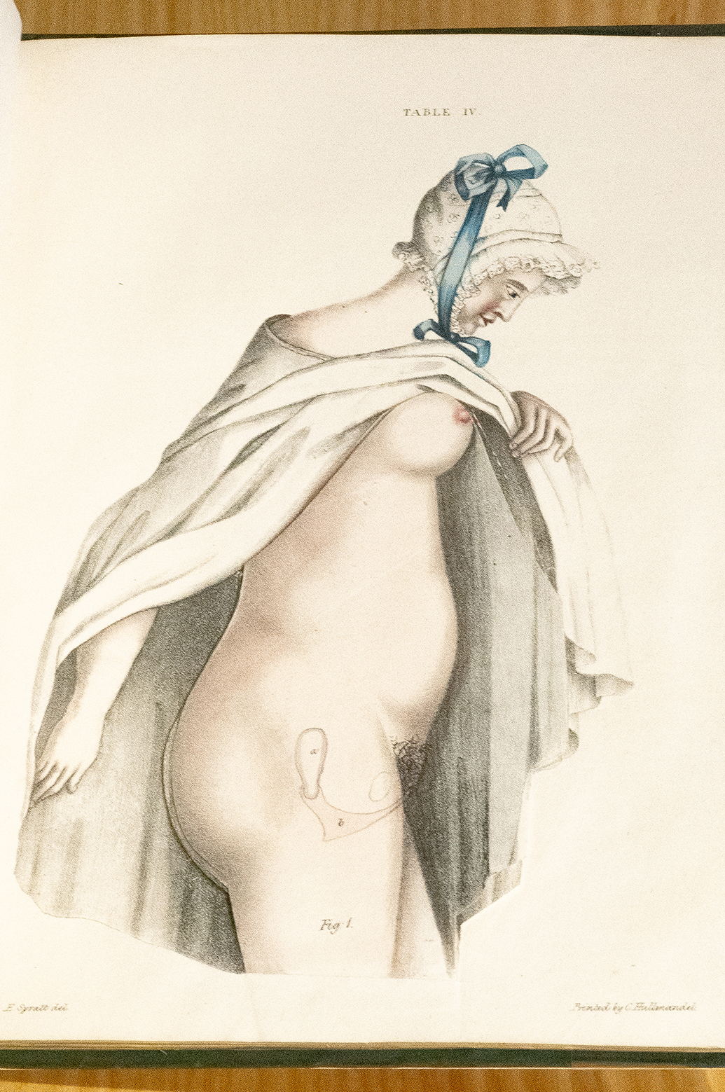
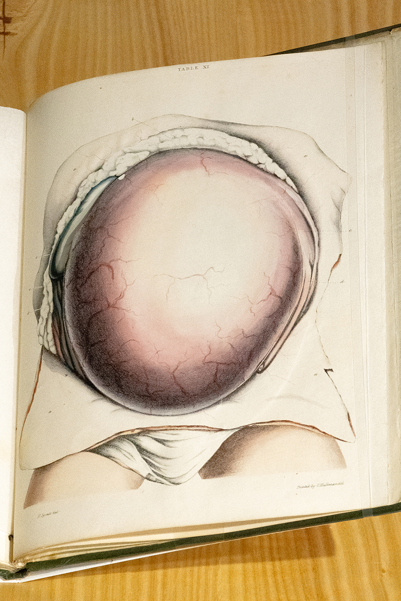
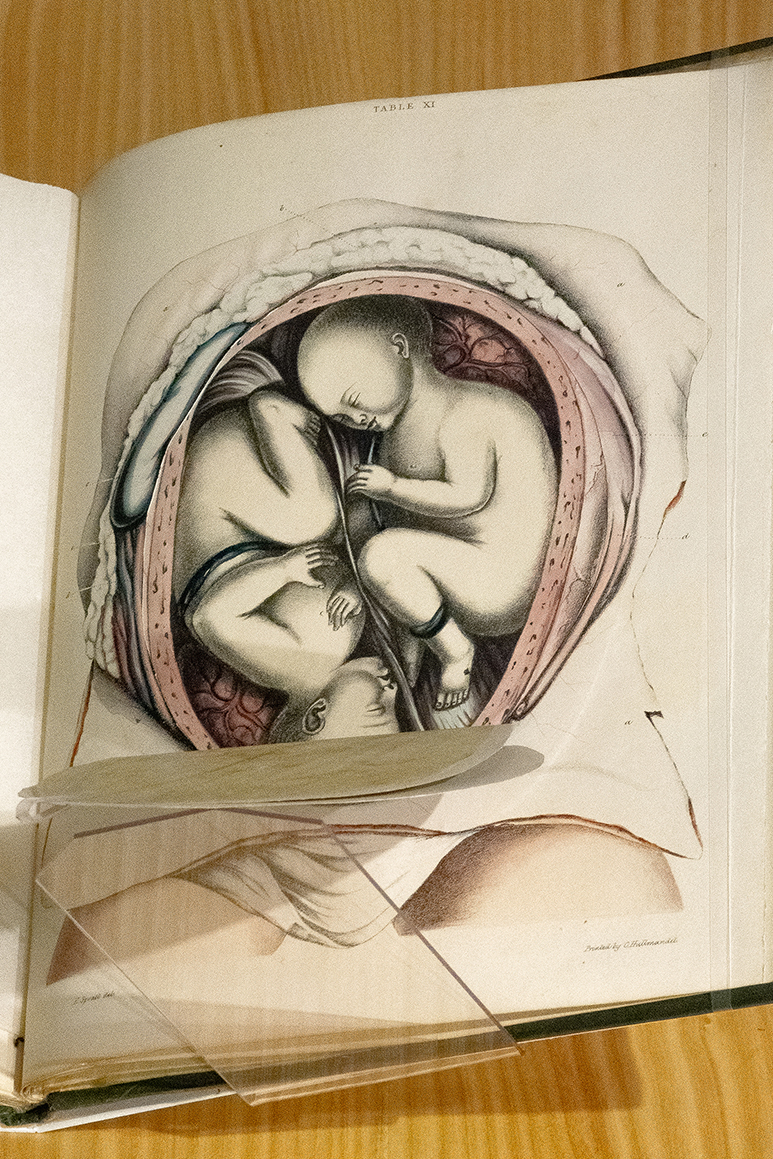
‘Supplement to obstetric tables’ (published 1835) by G. (George) Spratt
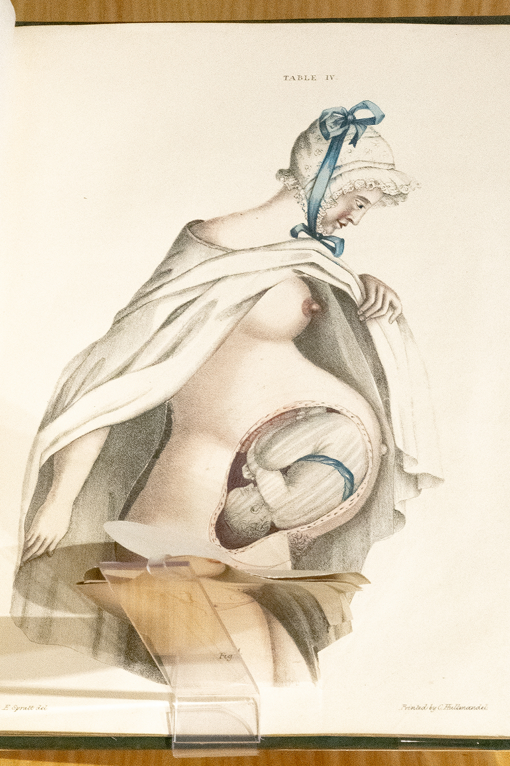
‘Supplement to obstetric tables’ (published 1835) by G. (George) Spratt
This striking flap anatomy in color prepared by the obstetrician George Spratt provides a glimpse into the uterus through a series of ‘dissections.’ Although intended for pedagogical use, this image invites reflection on the male medical gaze and its portrayal of the pregnant female body. On one hand, Victorian codes of sexual propriety and modesty are observed: the subject is wearing a frilly bonnet with blue ribbon and is holding up her pristine white shawl in order to facilitate her own anatomizing in the absence of intervening male hands. On the other hand, the very nature of the flap anatomy probing deeper into a vulnerable–because unclothed—female body suggests a voyeuristic aspect.
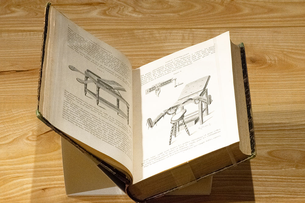
‘Traité de gynécologie opératoire’ (published 1885) by A. (Alfred) Hegar (1830–1914)
This image appearing in the French-language book by the German gynecologist, Treatise on Operative Gynecology, presents a reclinable table, complete with stirrups, for gynecological examinations and operations. The surrounding text provides details on the table’s dimensions and practical usage, suggesting that no detail has been spared in the male practitioner’s colonization of the space of the clinic and of the body of the female patient.
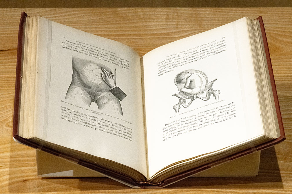
‘Traité du palper abdominal, au point de vue obstétrical’ (published 1889) by A. (Adolphe) Pinard (1844–1934)
These two images were intended to provide practitioners with techniques for ascertaining the fetal position in utero in the more dangerous instance of foot-first deliveries. The author Adolphe Pinard, a noted French obstetrician, was an advocate of obstetrical palpation, the technique of using one’s hands to ‘feel inside’ the body through the skin in order determine the position of the fetus. In the image to the left, a male practitioner’s hand easily perceives the fetal skull given its outward posture, while to the right, the fetal skull is hidden towards the back of the uterus.
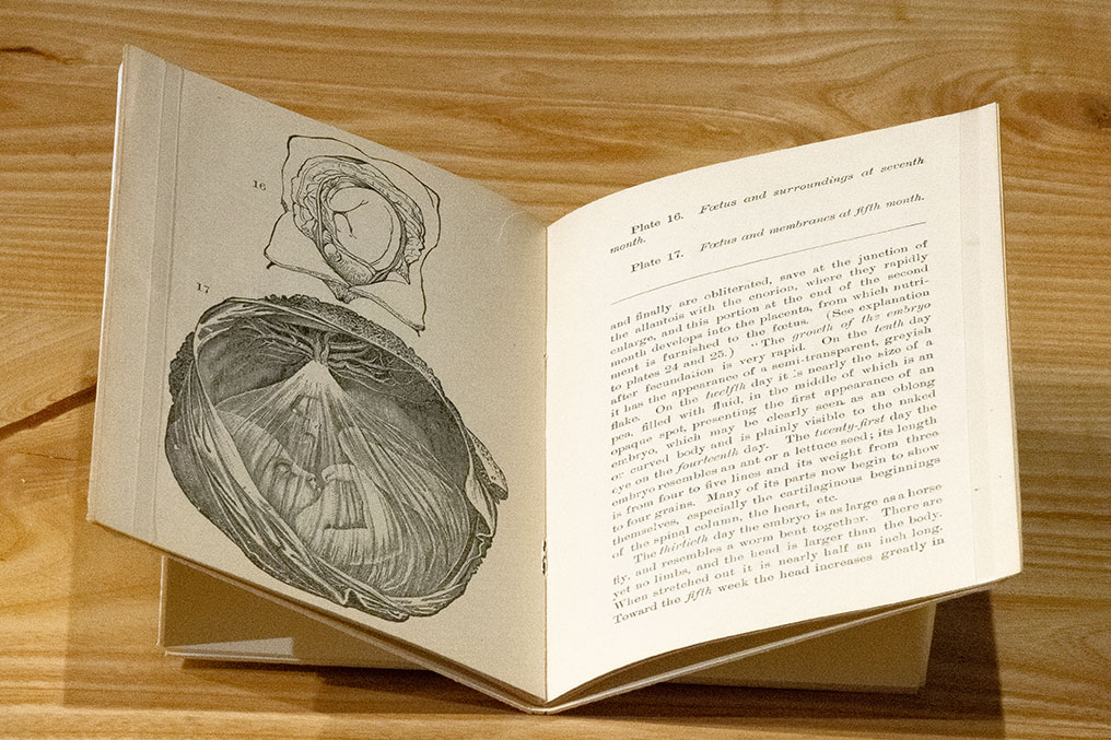
‘Maternity: a book for every wife and mother’ (published 1889) by P.B. (Prudence B.?) Saur
Here are two snapshots of the fetus in utero during its gestation: at six months (Figure 17) and seventh months (Figure 18) respectively. The accompanying text describes the shape and relative size of the fetus during the early days of its development. Use of the term “embryo” to describe the fetus at the thirtieth day points to the truly embryonic nature of its growth at a stage in which it has neither limbs nor a defined body, and rests in a seemingly delicate amniotic sac. The interchangeability of the terms “embryo” and “fetus” in this and other period textbooks suggests a different historical time period than our own. One in which the demarcation between the two did not have the political or legal consequences that it might today in the context of abortion, for instance.
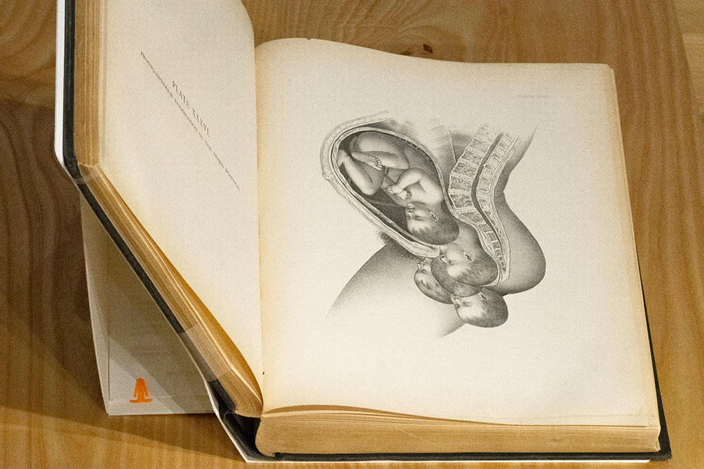
‘An illustrated encyclopaedia of the science and practice of obstetrics’ (published 1890) by F. H. Getchell, ed.
This striking lithograph plate from the Philadelphian physician F. H. Getchell’s obstetrical encyclopedia presents the side profile of a partially embodied mother, with a full-term fetus descending. The different positions of the fetal skull during delivery provide an animated effect that serve as a reminder of the dynamism and kinetic nature of the birthing process, as well as the male medical gaze’s approach to taxonomizing all the eventualities of childbirth.
The Fetus Dissected,
The Fetus in Folio
The Fetus Dissected,
The Fetus in Folio
The transformation of human anatomy into a scientific endeavor required new visuals that would capture and showcase the increasing complexity and medical value of the human body. Images of the fetus in utero have similarly evolved, taking prominence in the familiar or ‘classic’ anatomical texts that have contributed significantly to the fetus’ visual history. The materials in this case present the fetus in utero in the large-scale anatomy atlases known as ‘folios’ that were first popularized in the eighteenth century.
The fetus’ graphic entry into the folio tradition begins with a backstory: the re/turn to corporeal dissection in the late medieval period. The dissection of female corpses, particularly for the purposes of demystifying her organs of generation, was a prerequisite to studying, and then illustrating, the fetus in utero. In Antiquity, human dissection was largely forbidden, leaving Greek and Roman anatomists to dissect animals like Barbary apes, who were deemed suitable stand-ins for humans given their number of shared features. This prohibition on dissection contributed to the creation of anatomical images, including those of the fetus in utero, which were more imaginative than representative of reality.
From Antiquity to the Renaissance, there were several cultural and religious deterrents to bodily dissection. The most important of these was undoubtedly a Christian argument that the human body must remain intact for possible resurrection after death. For this reason alone, the dissectors of the Anatomical Renaissance like Vesalius went to great lengths to obtain corpses deemed suitable for dissection. “Suitable” bodies included persons who had been hanged or otherwise publicly executed for capital crimes, given that their moral transgressions on earth had already jeopardized their chances of salvation and resurrection. Even in these cases, certain municipalities required that corpses be retrieved from sites of execution outside the town limits, out of respect for the relatives of the deceased.
Thus Christian Europe had to become culturally comfortable with the medical uses of dissection before humans—let alone women—could be regularly opened up by anatomists for study. The normalization of dissection as a legitimate scientific activity was also facilitated by the rise of temporary scaffold structures that eventually became permanent, dedicated anatomy amphitheaters in Italian city states and the Netherlands, and then, across Europe. These venues were dedicated to teaching and research on the human body, functioning as an extension of the university Medical Faculty as the locus of innovation from which new images would emerge.
Opening up female bodies via dissection required another layer of care and attention given the broader culture of the strict segregation of the sexes outside of marriage. Physicians were not expected to touch patients outside of checking their pulse, for instance, leaving male anatomists to adopt techniques to combat potential charges of moral impropriety in handling female cadavers. One was to preserve the woman’s modesty by shrouding breasts and genitalia in textiles while other parts were being dissected—at least, this became a convention for anatomical teaching, both in situ and in textbook illustrations. Over time, the medicalization of the female body enacted by and through the male gaze reached a radical iconographic conclusion: that her body could be chopped and cleaved to reveal only the parts deemed most important or strictly necessary for obstetrical training. In this case, the mother’s sense of bodily integrity and personhood is compromised for the medical lesson at hand, as if dismembered “butcher shop meat” that must be sacrificed in service to the fetus.
The medicalized images presented here and elsewhere are open to further layers of interpretation. Many of those presenting mothers as merely reproductive vessels do so in evocative ways that betray a level of curation and staging that trespasses beyond mere pedagogical utility. Bare breasts primed for postpartum feeding, straight-on views of thighs spread apart for childbirth, and agonized-but-beautiful facial poses all hint at voyeurism. Indeed, there is potential for all such iconography to enter into the realm of sexually titillating or pornographic presentation. Underlying all imagery, whether taken as medicalized, sexualized, or some degree of both, is a notion of conventional female beauty that is encapsulated perhaps most clearly in the turn-of-the-twentieth-century life-size Pilz female “manikin.” She appears in full color with a period-appropriate lithe physique, symmetrical features, coiffed hair, and artful makeup. The tension between traditional representations of the female body and those of the pregnant mother as a scientific subject has often reconciled in favor of the fetus in utero, while nonetheless retaining traces of the maternal body.
The development and evolution of obstetric atlases and manuals aligned with a broader movement in medicine at this time that focused on consolidating its scientific culture and professional clinical practice. This progression embraced visuals that were stripped of artistic flourishes and presented standardized, yet realistic, images that conveyed clinically useful information. In terms of obstetric images, where the clinical focus shifted to the fetus, the depiction of the mother is rare and, when present, she often serves merely to orient the fetus within a corporeal geography. The mother has been increasingly measured and subsequently pathologized when she deviated from a “normal” morphology that best accommodates the fetus and facilitates safe and efficient childbirth. Similarly, images of the fetus in utero, while becoming more realistic, are presented in a way that stresses the dangers the fetus may encounter, often requiring mechanical intervention. Far removed from the spacious and nurturing uterus presented in earlier images, the fetus now appears more closely as it does in the ‘reality’ of our modern secular world. While created in the service of clinical objectives, these images represent a significant shift in the visual culture of the fetus in utero.
The Fetus Dissected, The Fetus in Folio
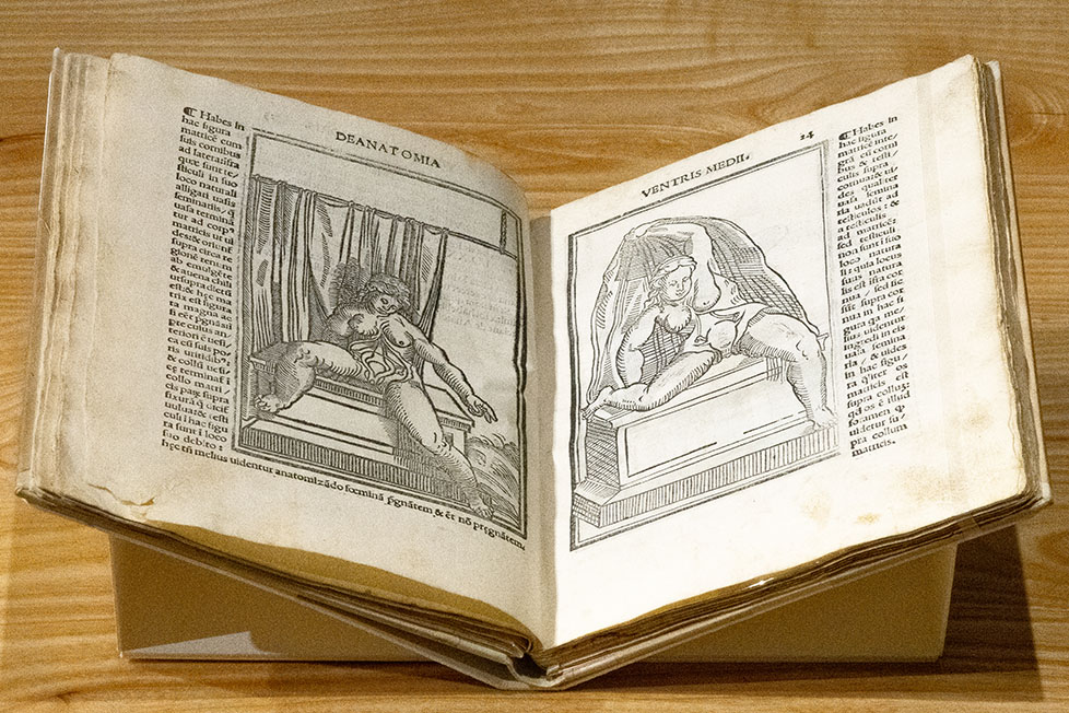
‘Isagoge breves prelucide ac uberime in anatomiam humani corporis’ (published 1522) by Jacopo Berengario da Carpi (c. 1460–c. 1530)
This two-page spread by the Bolognese-trained physician referred to as Carpus presents images of female organs of generation. Like most of the anatomy atlases of this vintage, the images are woodcuts, the text is in Latin, and the mother is presented fully embodied and adorned with artistic flourishes. In both, her organs of generation are on view, including what appear to be “uterine horns” in the right-hand image, in keeping with an early view that the uterus was configured in the shape of the devil’s horns.
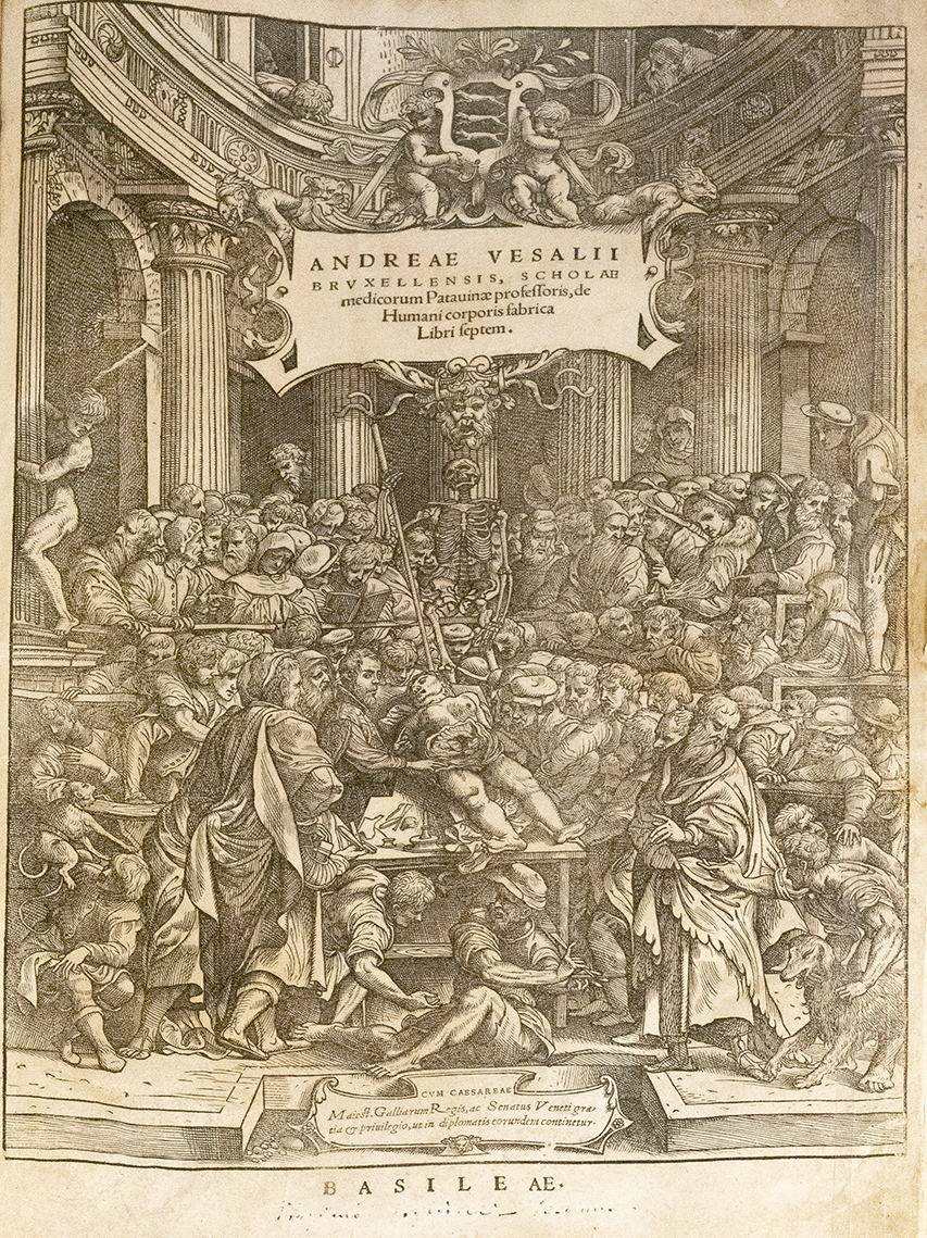
‘Scholae medicorum Patauinae professoris de humani corporis fabrica libri septem’ (published 1543) by Andreas Vesalius (1514–1564)
This elaborate woodcut frontispiece of the Latin-language Anatomy of the Human Body in Seven Books depicts its author, Andreas Vesalius, dissecting a female cadaver surrounded by a throng of students in an early makeshift anatomy theater. The corpse was that of a hanged criminal who had attempted to stay her execution by claiming she was with child. After calling in a midwife to ascertain that the condemned woman was not in fact pregnant, her execution was carried out, and her body was anatomized for the sake of furthering Vesalius’ agenda to arrive at new knowledge through corporeal dissection. It is believed that the midwife whose examination led the criminal to her death is one of two living female figures depicted here. Most likely, she is the cloaked figure directly above the handrail to the left of the central skeleton who is casting a pained, perhaps guilt-ridden, glance at the cadaver. Vesalius’ decision to showcase a female corpse on the cover of his masterpiece reveals the cachet of obtaining women for dissection at a time when sourcing corpses of either sex was by no means easy.
‘La dissection des parties du corps humain’ (published 1546) by Charles Estienne (1504–ca. 1564)
Charles Estienne was a French anatomist of the early Renaissance, at a time when the ancient practice of dissecting corpses was being revalued as an indispensable aspect of the anatomist’s work. Although Estienne trained under the ancient Galenic orthodoxy, he used the knowledge gained from hands-on dissecting to question traditional views and further new anatomical research. This image presents a fetus in utero that has seemingly been unfurled as if a scroll. His flayed mother presents him stoically in an open air urban setting that is most unusual for anatomical depictions in this period, which typically favor naturalistic settings.
‘Vivae imagines partivm corporis hvmani aereis formis expressae’ (published 1566) by Juan Valverde de Amusco (c.1525–c.1588)
This copperplate engraving appears in a book featuring the “living images” of parts of the human body. Although its author was an adversary of Andreas Vesalius, the greatest living anatomist at the time, this image is typical of Renaissance anatomy. It presents a female statute-like figure or Venus who is stoically performing the impossible feat of presenting her own flayed abdomen, for all to see. She appears in a naturalistic setting and is flanked by two sets of additional images, with the bottom right featuring both a fetus in utero and ex utero.

‘De humani corporis fabrica librorum epitome’ (published 1642) by Andreas Vesalius (1514–1564)
The most famous of the Renaissance anatomists, Andreas Vesalius gained notoriety as a hands-on dissector who wished to correct the knowledge of ancient anatomists. In this frontispiece of the Epitome, a companion piece to his path-breaking 1543 publication Anatomy of the Human Body in Seven Books, a female corpse is also at the center. Although in the revival of human dissection the male body was taken as the measure of all things, anatomists like Vesalius increasingly turned their attention to the peculiarities of the female body, especially the gravid uterus. It follows that a precondition to unveiling the fetus in utero was to uncover the female body, who was transformed into a subject of study in her own right. In this allegorical woodcut, dissectors have opened the abdomen of a recumbent woman whose head is shrouded in mystery—a technique adopted to preserve her anonymity.
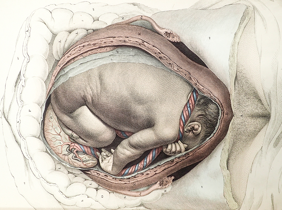
‘Anatomia hvmani corporis, centum & quinque tabvlis, per artificiosiss’ (published 1685) by Govard Bidloo (1649–1713)
Govard Bidloo was an anatomist of the seventeenth-century Dutch Golden Age. As a playwright, poet, and physician, Bidloo emblematized the burgeoning nation in his dynamic approach to human anatomy. His naturalistic graphic approach captures something of the everyday interior or “genre” tradition that flourished in Dutch painting, in which the “burgher” or middle merchant class sought to adorn their homes with artworks representing their world. But of course, the subject of this engraving is anything but “everyday.” Here, the viewer is confronted with a partially dissected, nude female corpse with flayed abdomen exposing a near-term fetus and placenta in utero.
‘The anatomy of humane bodies, with figures drawn after the life’ (published 1698) by William Cowper (1666–1709)
The publication of this work by the English surgeon-anatomist William Cowper gained him great acclaim and a prestigious membership to the Royal Society, the first state-funded scientific institute in the Western tradition. Among others, this engraving features a deceased eight-month-old male fetus ex utero who remains attached to the placenta by the umbilical cord.
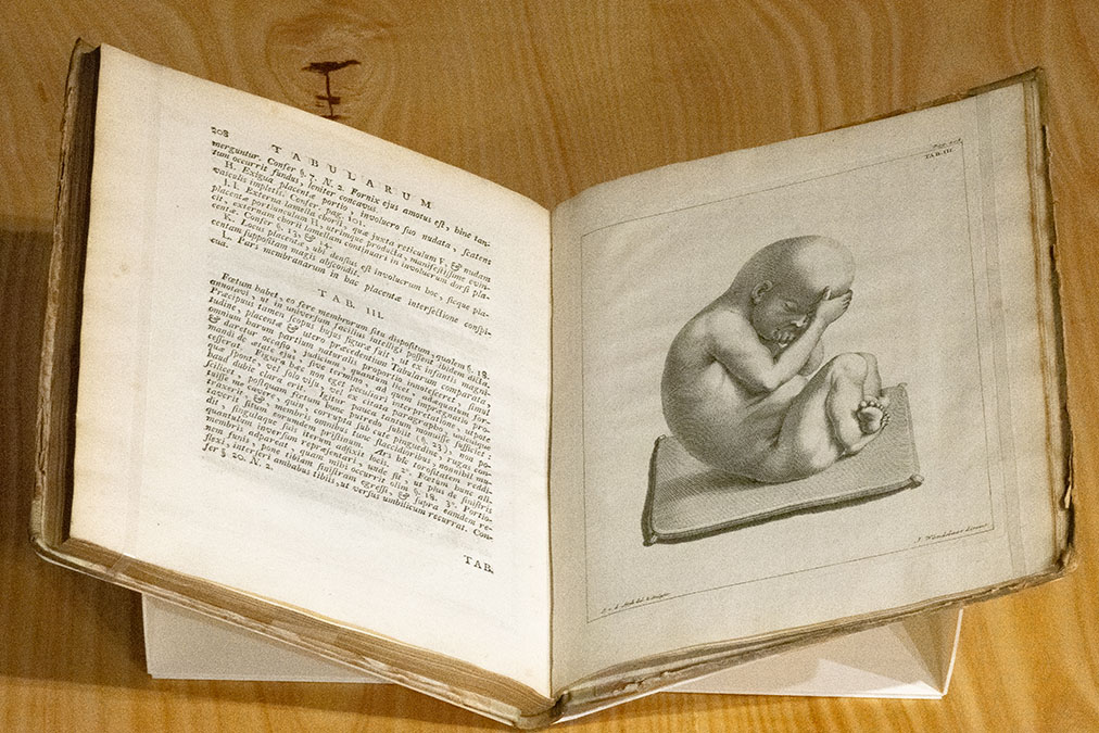
‘Uteri humani gravidi anatome et historia’ (published 1743) by Wilhelm Noortwyk
This Latin-language text, Anatomy and History of the Gravid Human Uterus, was published by the Dutch anatomist Wilhelm Noortwyk (or Noordwyk). This engraving captures the fetus entirely ex utero, seated on a thin mat, suggesting its posthumous removal from a female corpse for examination.
‘Tabulae anatomicae’ (published 1758) by William Smellie (1697–1763)
This engraving portrays the growing fetus as a mature being—the early-term fetus in Figure 1 already has pronounced facial features, while the Figure 2 fetus has abnormally developed phalanges and strikingly defined musculature. The mother is entirely absent from these engravings, as if she plays a limited role in the generative process. She is merely represented by her pelvis—cumbersome bones seen by male midwives as an interference to successful labor. William Smellie’s tools, represented in other images, were often perceived as rescuing the full-term fetus in utero from the woman’s restraining pelvis during the delivery process. Caption written by Amber Olson.
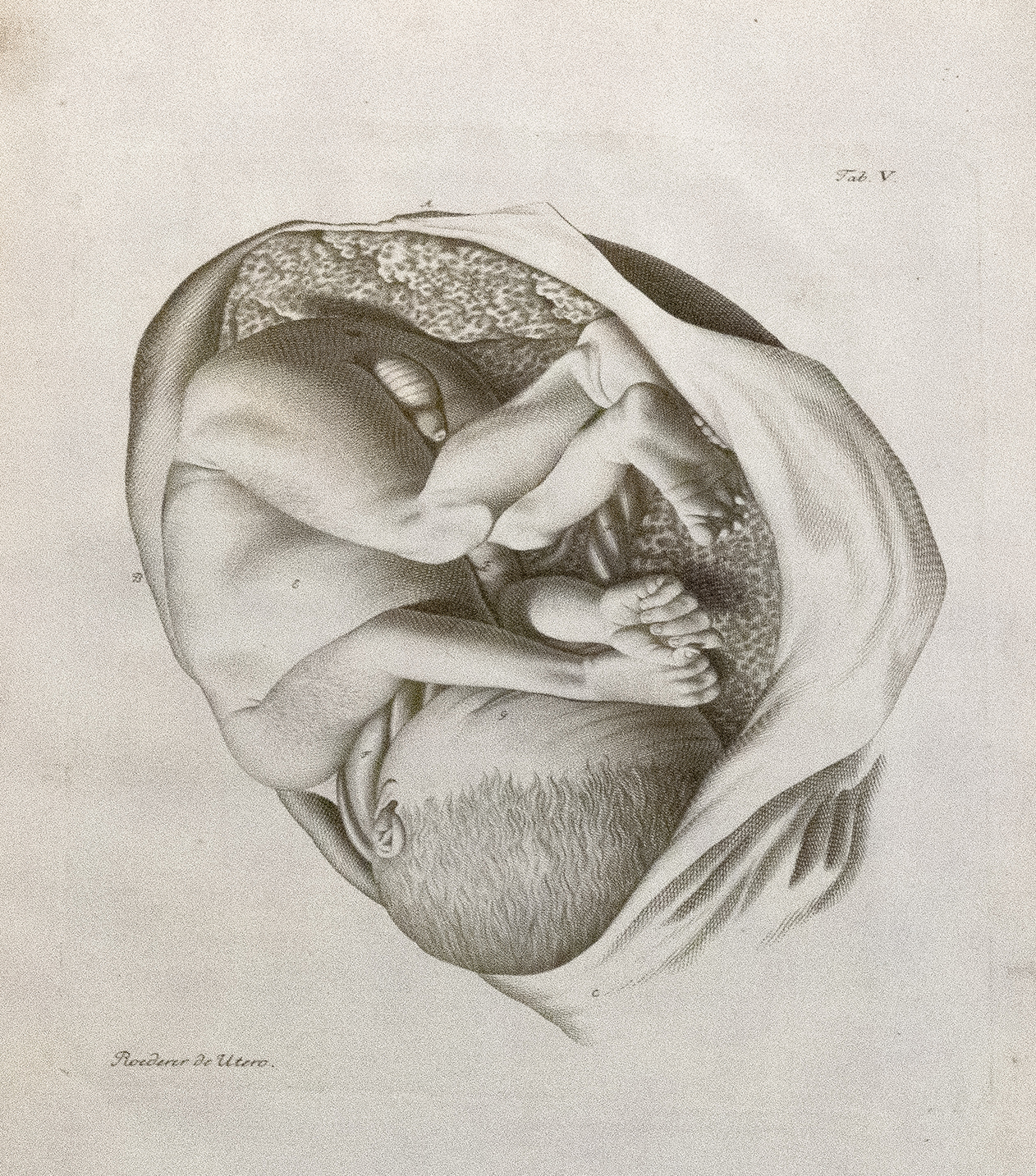
‘Icones uteri humani observationibus illustratae’ (published 1759) by Johann Georg Roederer (1726–1763)
This anatomy atlas by the German physician and professor of obstetrics at the University of Göttingen, Johann Georg Roederer, presents the fetus in a flayed uterus, indicating it has been copied from a dissection of a deceased subject.
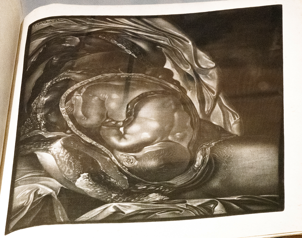
‘Abbildung der Gebähr-Mutter aus einer schwangern Frau’ (published 1761) by Charles Nicholas Jenty (?–at least 1777)
This striking mezzotint engraving features in the work of the French surgeon and anatomist Charles Nicholas Jenty. The sophisticated use of black, white, and shading renders an almost photocopier quality image of a unique, if gruesome, view onto the flayed cadaver of a deceased female patient who had carried the child to term.
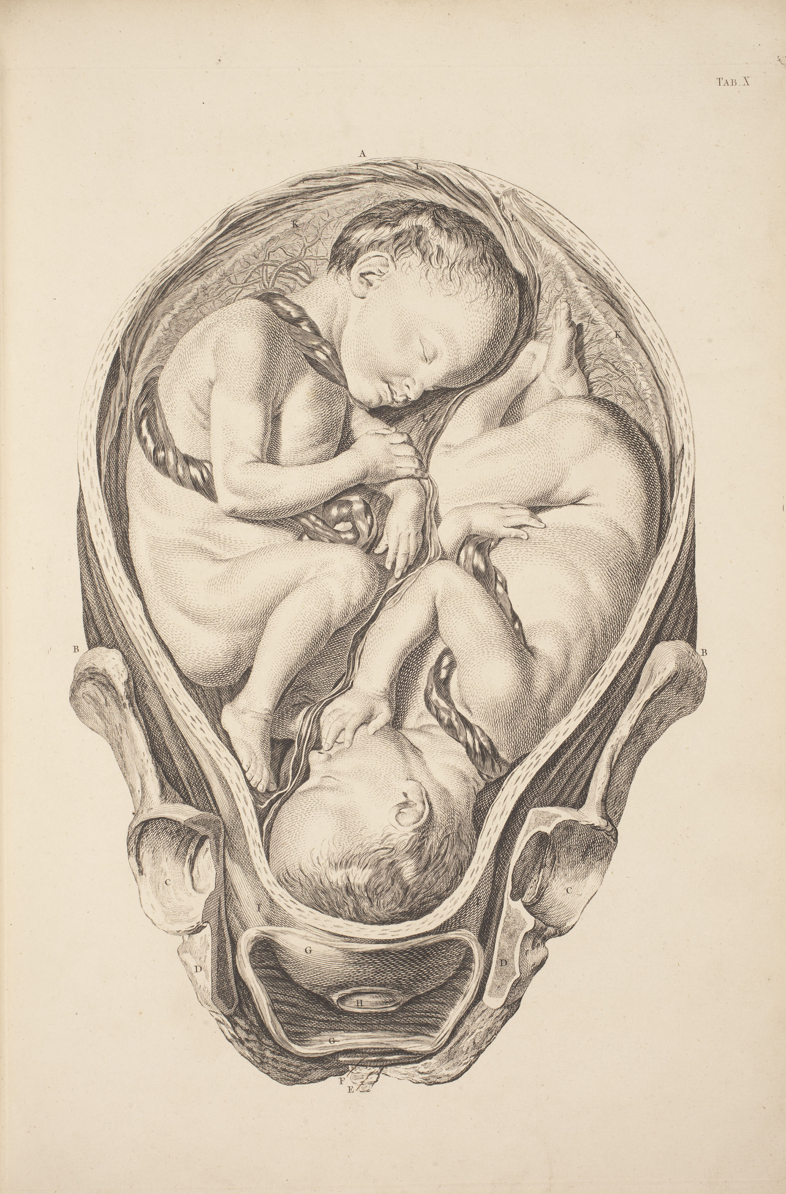
‘A set of anatomical tables, with explanations, and an abridgment, of the practice of midwifery’ (published 1761) by William Smellie (1697–1763)
William Smellie’s Anatomical Tables showcase the Scottish man-midwife’s commitment to teaching childbirth and his development of controversial technologies like the obstetrical forceps, which he argued made for safer deliveries. Although no such instruments are present in this image of twins in utero, his selective presentation of the mother’s womb encased by the bony pelvis suggests a mechanical approach to childbirth focused on the potential obstructions occasioned by a narrow passageway.

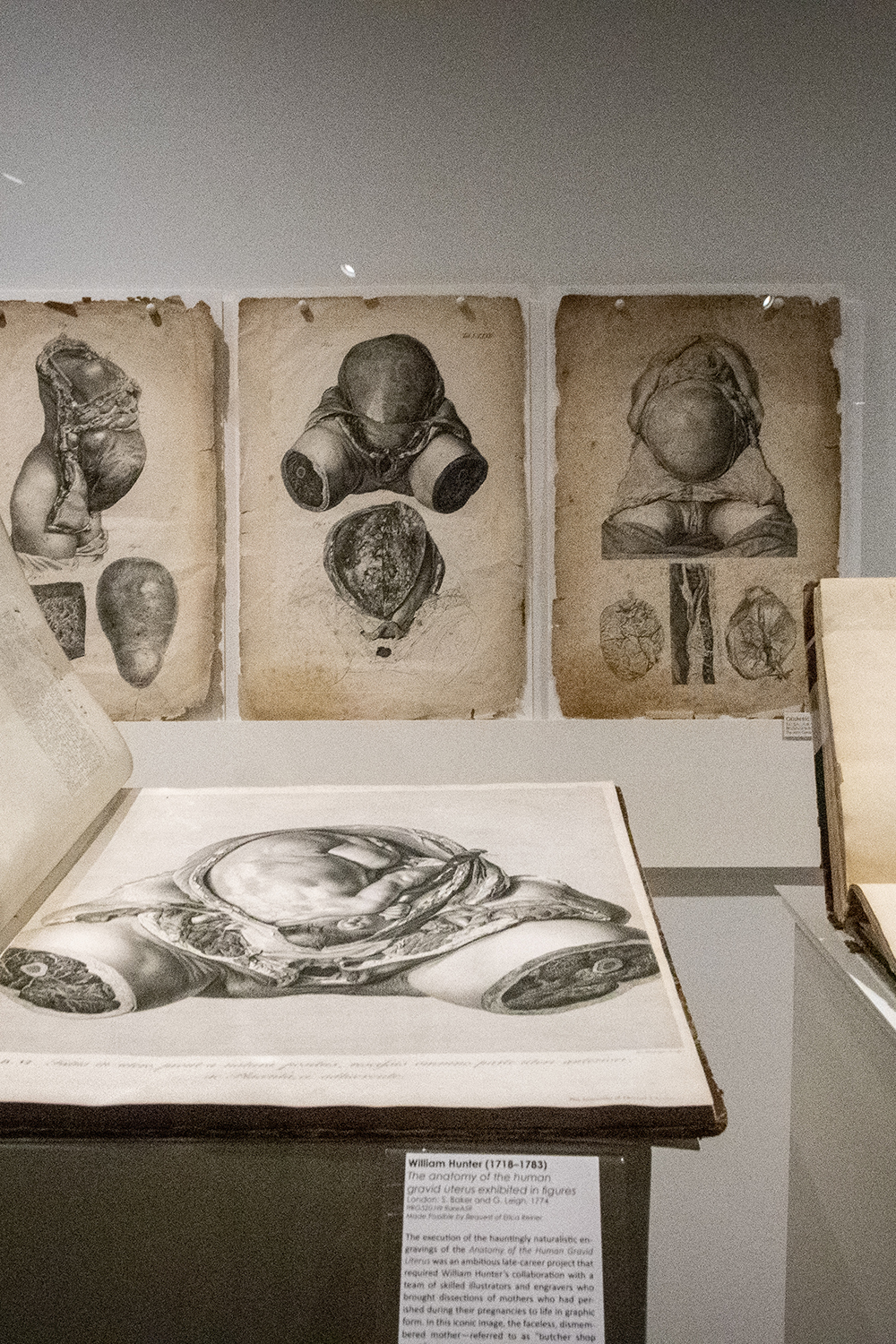
‘The anatomy of the human gravid uterus exhibited in figures’ (published 1774) by William Hunter (1718–1783)
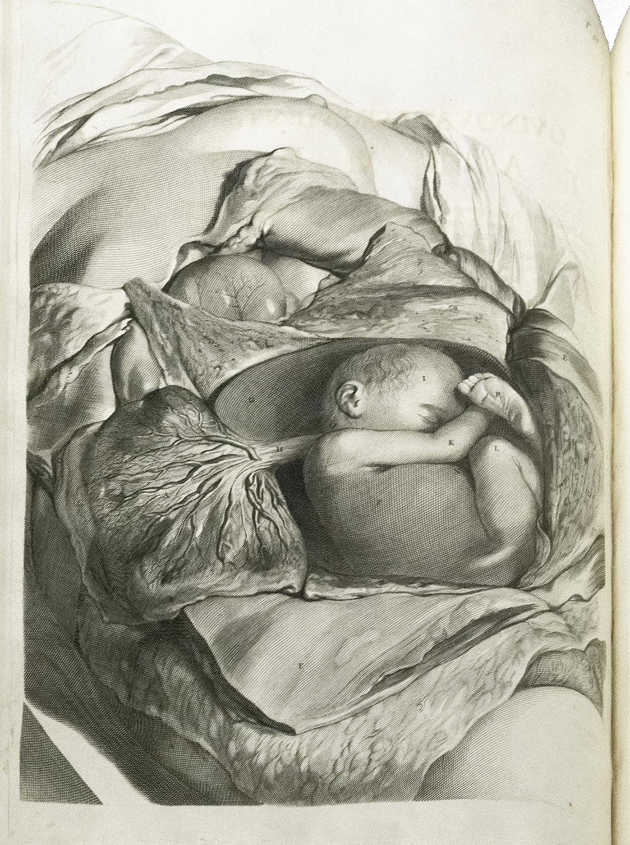
The execution of the hauntingly naturalistic engravings of the Anatomy of the Human Gravid Uterus was an ambitious late-career project that required William Hunter’s collaboration with a team of skilled illustrators and engravers who brought dissections of mothers who had perished during their pregnancies to life in graphic form. In this iconic image, the faceless, dismembered mother—referred to as “butcher shop meat”—is carrying a near-term fetus in utero.

‘The anatomy of the human gravid uterus exhibited in figures’ (published 1818) by William Hunter (1718–1783)
Over the course of his career running the Anatomy School on Great Windmill Street in London’s Soho district, William Hunter dissected hundreds of bodies with his students. This collaborative experience primed him for the completion of the most ambitious project of his career on the gravid human uterus. This remarkably naturalistic engraving depicts a full-term fetus primed for delivery in the head-first position. The apparent, but yet heavily curated, realism reveals not only the skill of the illustrators and engravers behind the images, but also the attention to detail sought by Hunter in executing this work.
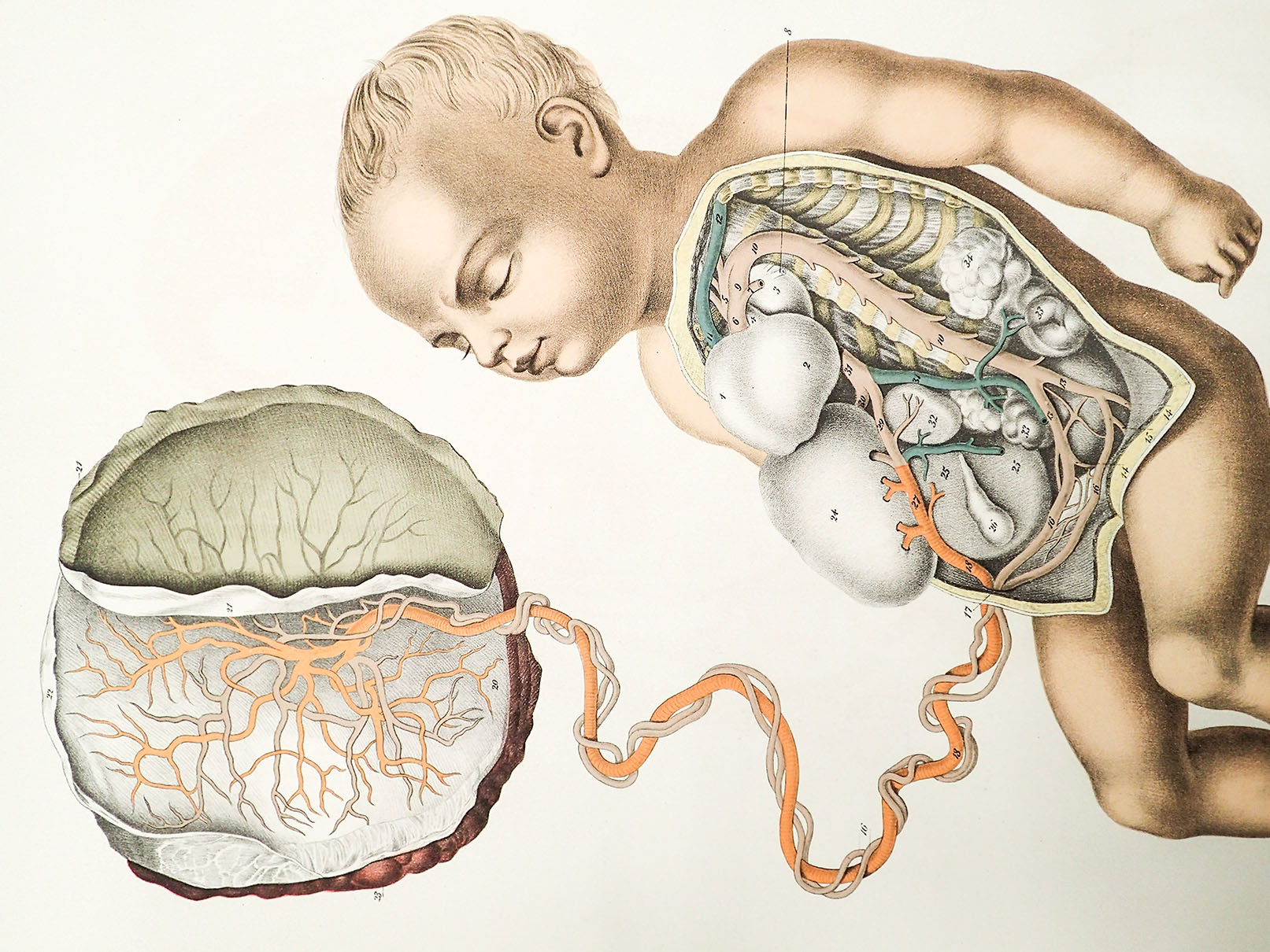
‘The Anatomy of the Bones of the Human Body’ (published 1832) by Jean-Joseph Süe (1710–1792
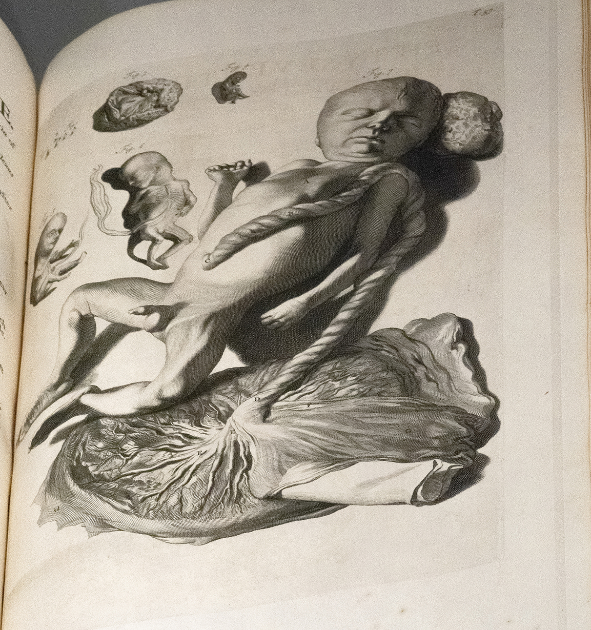
In this image, the fetus is entirely unfettered, with the exception of its umbilical cord and placenta. These parts have been isolated and the fetus has been dissected out of the uterus, in order to portray the interaction between fetal and maternal blood vessels, colored in blue and red.
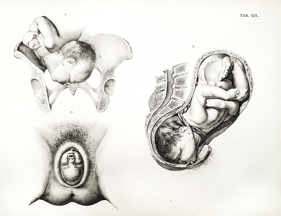
‘Geburtshülflicher atlas in 48 tafeln und erklärendem texte’ (published 1835-44) by Hermann Friedrich Kilian (1800–1863)
Hermann Friedrich Kilian was a German physician who, like many of his generation, travelled to other European anatomy schools—those in London, Paris, Vienna, and St. Petersburg—to further his education. This Obstetrical Atlas of forty-eight plates with explanatory notes showcases the fetus in utero disembodied from its mother in ways revelatory of the male medical gaze. Here, three shockingly realistic images present the practitioner’s concern over fetal-maternal skeletal interactions and birthing positions.
‘A system of anatomical plates of the human body’ (published 18–) by John Lizars (1787?–1860)
John Lizars was one in a long line of renowned Scottish anatomists who specialized in such subjects as ovarian disease. This color lithograph plate depicts a full-term fetus in utero that has been partially ‘dissected out’ of its disembodied mother, whose abdomen has been cut open. Though her external genitalia are on display, a white sheet suggests that she is being handled with modesty in mind.
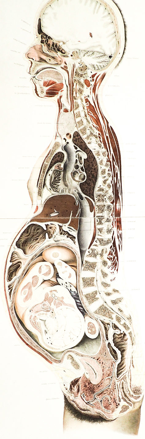
‘Die lage des uterus und foetus am ende der schwangerschaft’ (published 1872) by Wilhelm Braune (1831–1892)
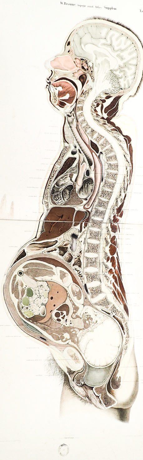
‘Die lage des uterus und foetus am ende der schwangerschaft’ (published 1872) by Wilhelm Braune (1831–1892)
This cross-section of a pregnant woman’s body depicts and labels her internal organs and presents a full-term fetus in utero. Unlike the dissected mother, the fetus’ entire body remains intact—complete with facial expressions—juxtaposing fetal humanity with her scientific presentation. The chart’s cross-sectional perspective reveals a tension between the scientific objective of probing the body and the maternal vulnerability that this male medical gaze reinforces. On one hand, this perspective serves an important clinical function by defining positional relations of structures relevant to surgery. While of questionable clinical utility at the time, such cross-sections of the fetus in utero are prescient precursors of the fetal ultrasound and imaging modalities like the CT and MRI, which have become the dominant image of the fetus in utero. On the other hand, this perspective has separated the mother from the reproductive process by the two-part act of de-feminizing and then re-masculinizing her anatomy. It appears that an ‘artistic mastectomy’ has been performed, with Braune chopping off any evidence of her breasts and exhaustively labelling all organs except for her genitalia. The short hairs around her skull, in turn, mimic a typical male hairstyle or that of a young child. Caption written by Madeline de Figueiredo.
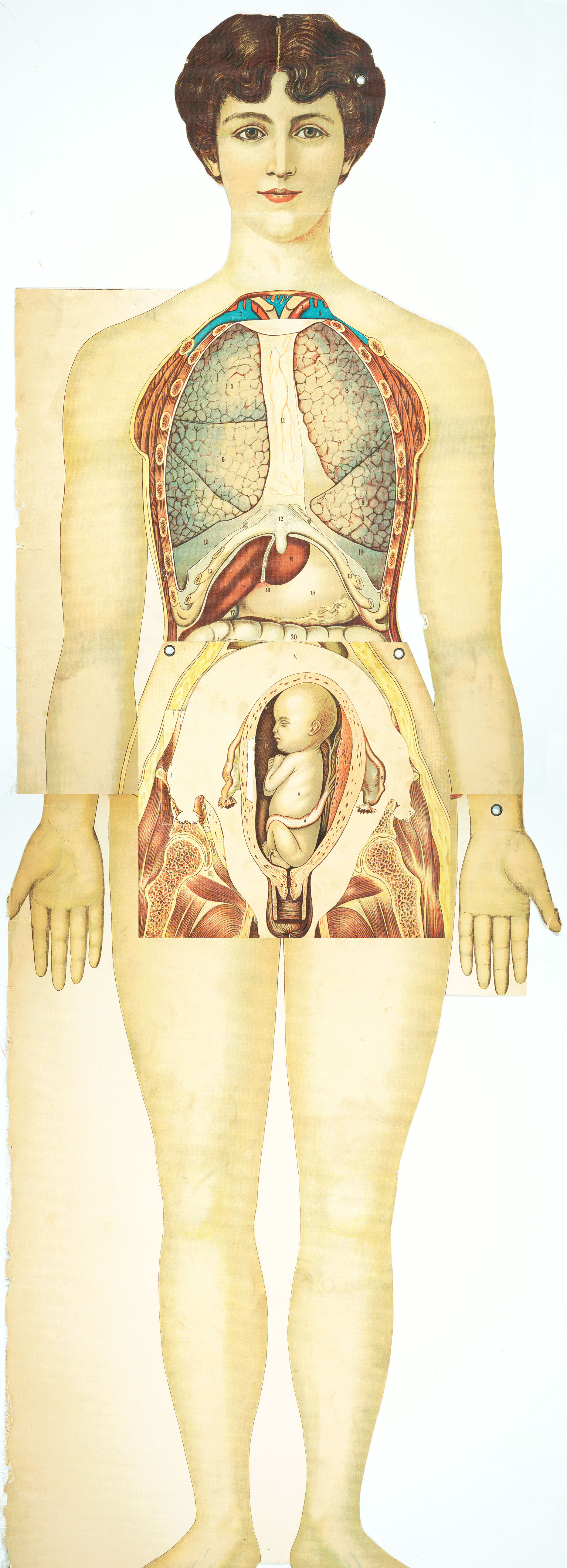
‘Pilz anatomical manikin [female]’ (published 19–)
This striking lifesize female manikin in full color served as a pedagogical tool for turn-of-the-twentieth-century medical students. Her stylized and aesthetically oriented features—including such touches as coiffed hair and makeup—reveal contemporary notions of female beauty and the “ideal” shape, which in this period was a lithe physique, in contrast to the more voluptuous Renaissance Venuses. She is nonetheless presented with a healthy ‘maternal’ hip-to-waist ratio. The manikin is designed to be used to display the human skeletal, muscle, and nervous systems, as well as other internal organs, which can be added to and subtracted at will, with clever flaps and overlays. The overlay depicted here presents the fetus at various stages of development, culminating in full term. The baby presented is healthy and robust, with a full head of hair.
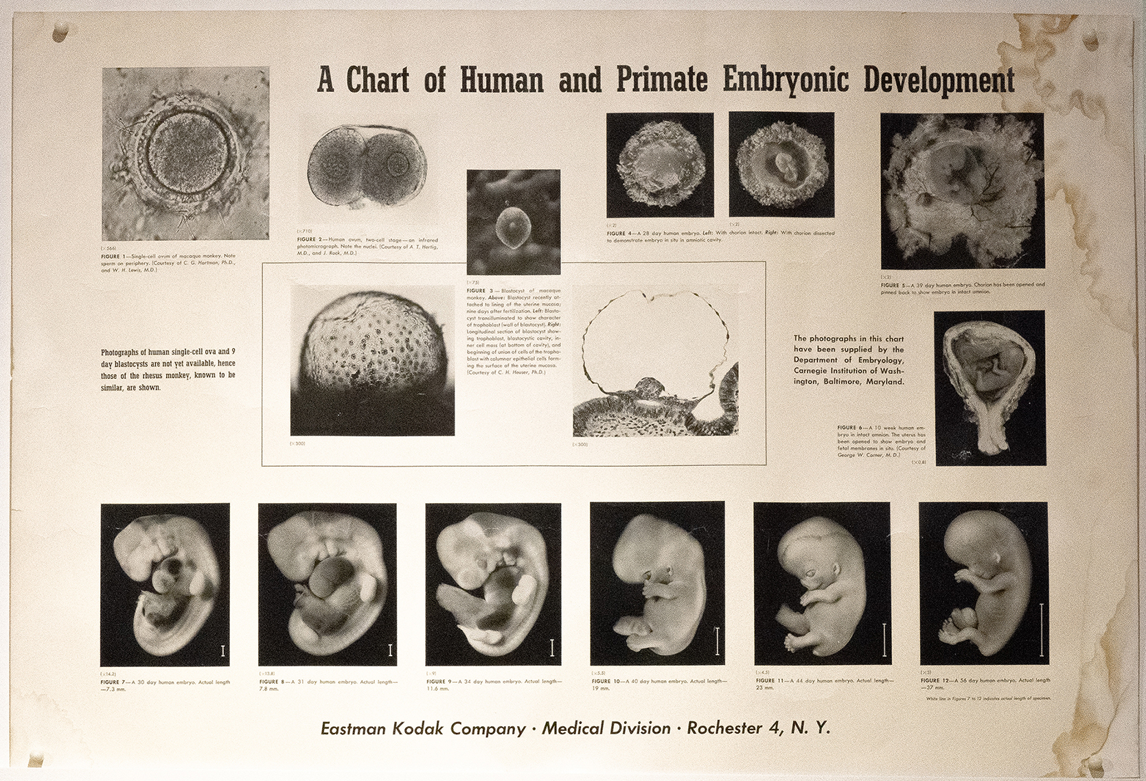
‘Medical School Anatomy Labs development chart’ (published n.d.)
This educational chart, relying on the objectivity of photography, presents fetal development in a concise scientific narrative.
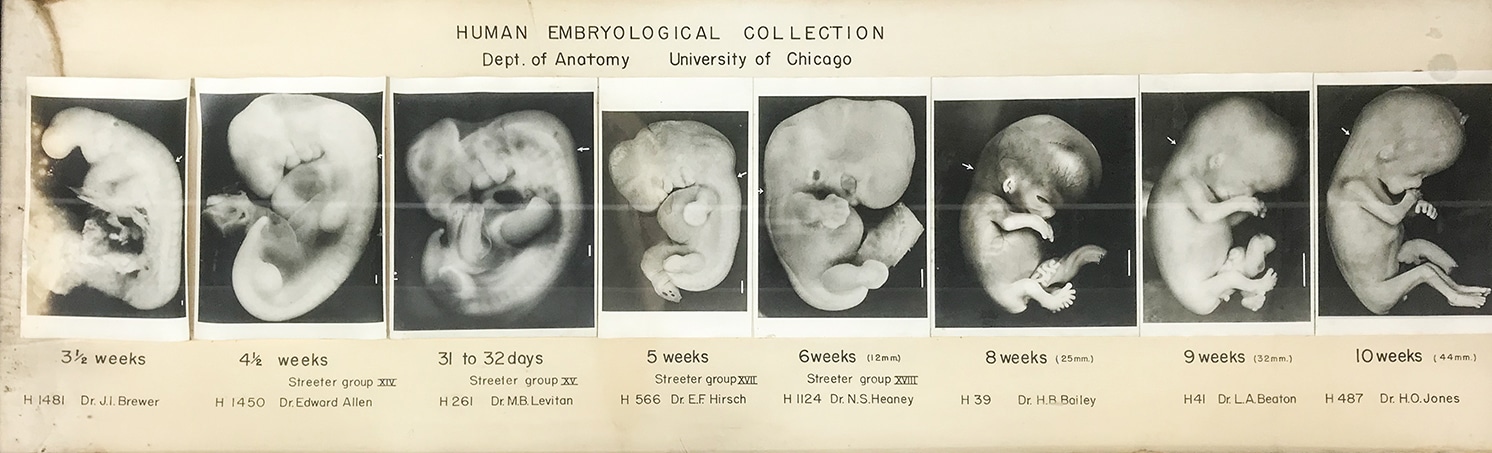
‘Medical School Anatomy Labs human embryological collection’ (published n.d.)
Photography and the Fetus
Photography and the Fetus
Photography as a technology that “captures” reality is often considered objective, and for this reason, has been readily adapted to medicine and use in anatomical and clinical texts. However, photographing human tissue in black and white was technically challenging, often resulting in images of an indistinct mass of tissue, blood vessels, and organs, as some of these images attest. Additionally, early photographic technology was limited in what it could see; it did not yet allow for visualization of the fetus in utero. As a result of these limitations, early obstetrical photographs often depicted deceased mothers and fetuses in various stages of dissection, resulting in images not palatable for dissemination beyond the medical domain. The voyeuristic concerns of obstetrics and the harshness of the medical gaze were further called into question by the early use of photography in obstetrics.
Photographs like those found here of anonymous—and perhaps unwitting and unwilling— female patients raise questions about the changing standards of patient consent and the powerful position of male medical authority in this period. The violence done to the body is clear and invites a modern-day feminist critique of female erasure. Not only is the mother often removed from these images, what remains of the female body is reduced to its most useful fragments and bones relevant to reproduction—the uterus and pelvis. However, these images also suggest that the evolution of obstetrics at the hands of male practitioners was premised on notions of ‘progress’ that privileged some viewpoints over others. In this case, the mother’s erasure from decision-making in childbirth is treated merely as the collateral damage of the longer arc towards transparency in fetal representations.
Owing to both its content and the novelty of stereoscopic technology, The Edinburgh Stereoscopic Atlas of Obstetrics makes a unique contribution to the visual culture of the fetus in utero. Comprised of 100 images, published in four volumes between 1908 and 1909, this atlas adopts the entertainment aspect of stereoscopy with the obstetrical medical gaze, resulting in a visual spectacle that both captivates and repels. The morbid specimens in these images come to life through the illusion of 3D. Forced to view through the stereoscope to achieve the 3D effect, the viewer’s gaze is directed at images that are often gruesome and bordering on illicit. While the display of the anatomical specimens in the atlas serve as photographic updates of many of the graphic representations seen in prior eras, viewing these images as intended invokes a sense of voyeurism. While presented as an educational object, the utility of this atlas, beyond its novelty, has been questioned. It nonetheless remains a pioneering atlas containing some of the first published photographs of a live childbirth.
While the technology of early photography was limited in its ability to capture the fetus in utero and the images that were created were primarily confined to the medical domain, subsequent developments in photography significantly altered the visual culture of the fetus in utero. These modifications created an opportunity for these images to spread far beyond the traditional medical and educational domains. This new era in the visual culture of the fetus in utero began in April 1965, with the publication of Lennart Nilsson’s now iconic photographs in Life magazine.

Photography and the Fetus

Life Magazine, April 30, 1965 cover by Lennart Nilsson
This Life magazine cover photograph depicting a fetus in utero quickly gained iconic status after its publication on 30 April 1965, with eight million copies selling within four days of its release. The fetus is disembodied from its mother and appears to float freely through space and time, as if to anticipate the astronauts who would colonize the moon four years later and secure American superiority over the Soviet Union in the infamous “Space Race” of the Cold War. The photograph was one of a series of career-making images for the journalist, Lennart Nilsson (1922–2017), who captured fetuses like these in the context of ongoing abortion debates in his native Sweden.
Although later in life, he appeared agnostic on the abortion question, at the time this and other images were published in his book A Child is Born (1965), Nilsson was enthralled by the pro-life physician, Per Wetterdal. It was through Wetterdal that Nilsson gained access to the first fetuses he ever photographed at a Stockholm Women’s clinic. While these images proved pivotal in debating new abortion legislation in Sweden at the time, they were presented in America less controversially, as examples of the wonders of the human biological world. What very few if any consumers of this iconic fetal image knew then, as now, was the degree of curation required to produce this healthy looking fetus: Nilsson used backlighting and other photographic techniques to present the fetus as if alive, where in fact he primarily photographed aborted fetuses.
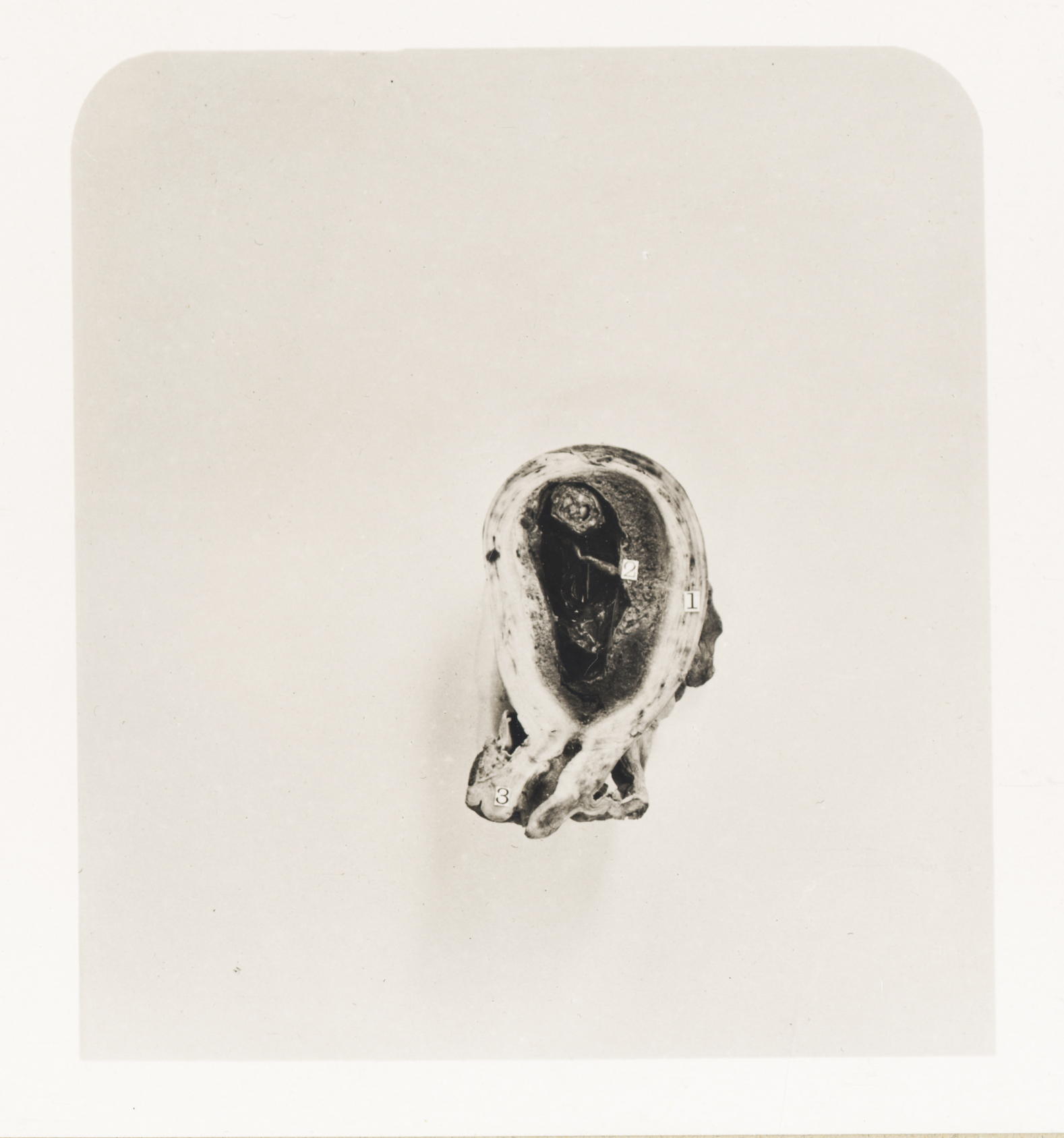
‘Gravid Uterus at Two and a Half Months’ from The Edinburgh Stereoscopic Atlas of Obstetrics (published 190-) by G.F. Barbour Simpson
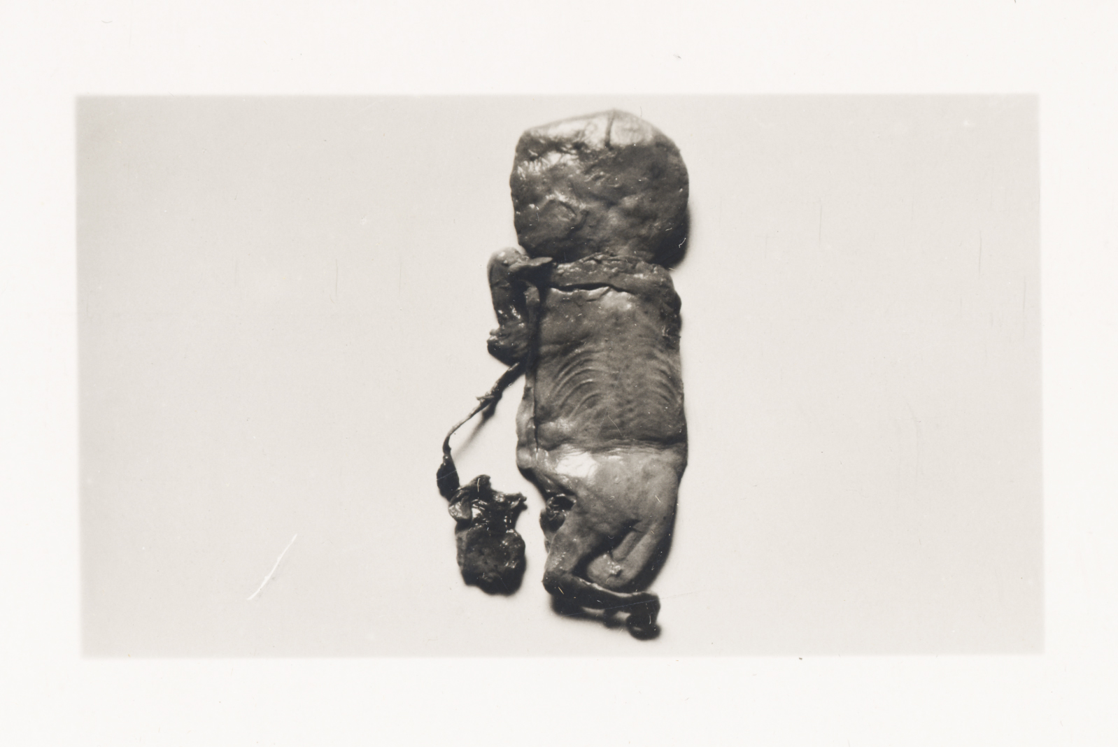
‘Foetus Papyraceus’ from The Edinburgh Stereoscopic Atlas of Obstetrics (published 190-) by G.F. Barbour Simpson
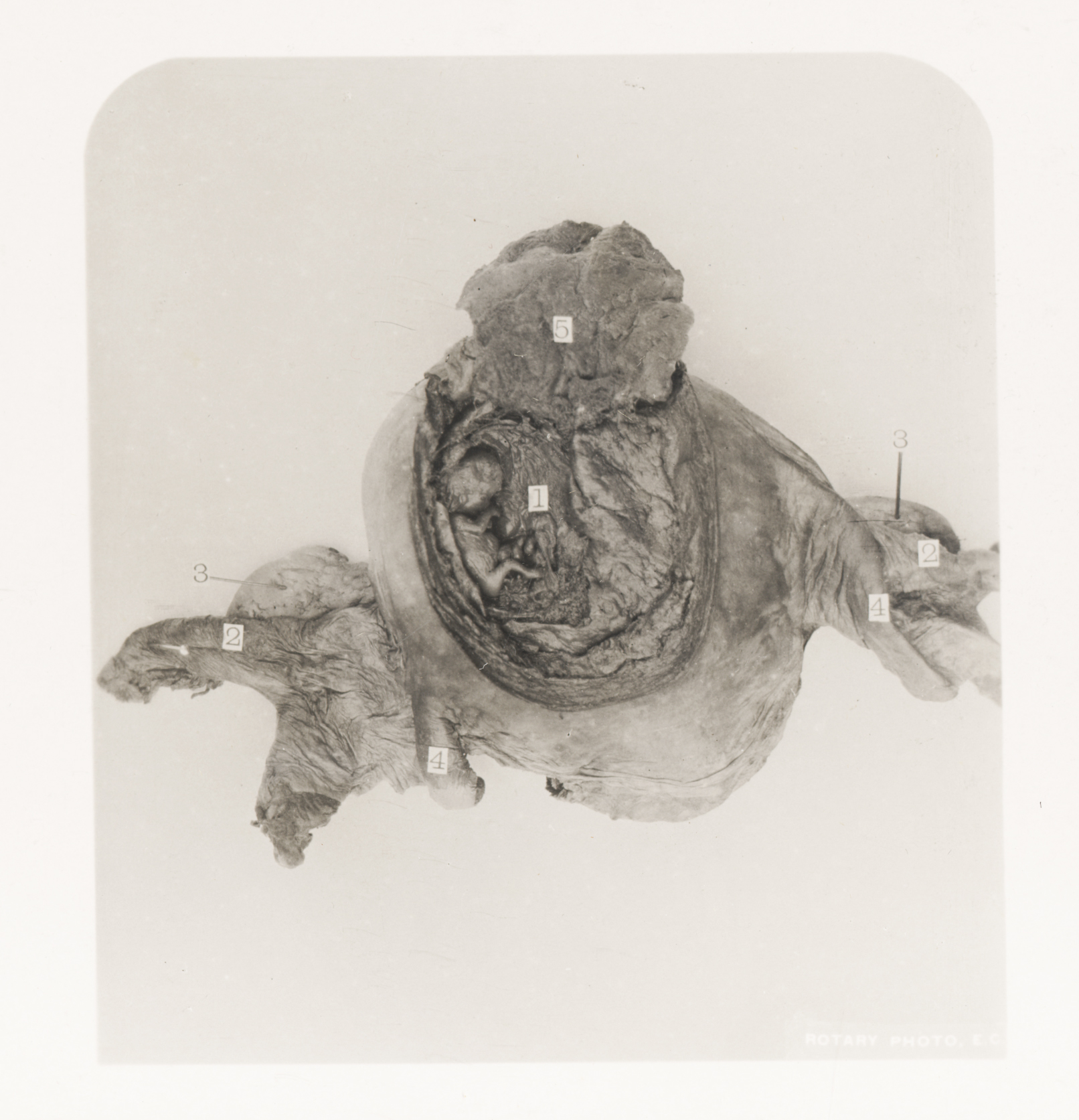
‘Gravid Uterus at the Third Month of Pregnancy’ from The Edinburgh Stereoscopic Atlas of Obstetrics (published 190-) by G.F. Barbour Simpson

‘Gravid Uterus at the Eighth Month of Pregnancy’ from The Edinburgh Stereoscopic Atlas of Obstetrics (published 190-) by G.F. Barbour Simpson
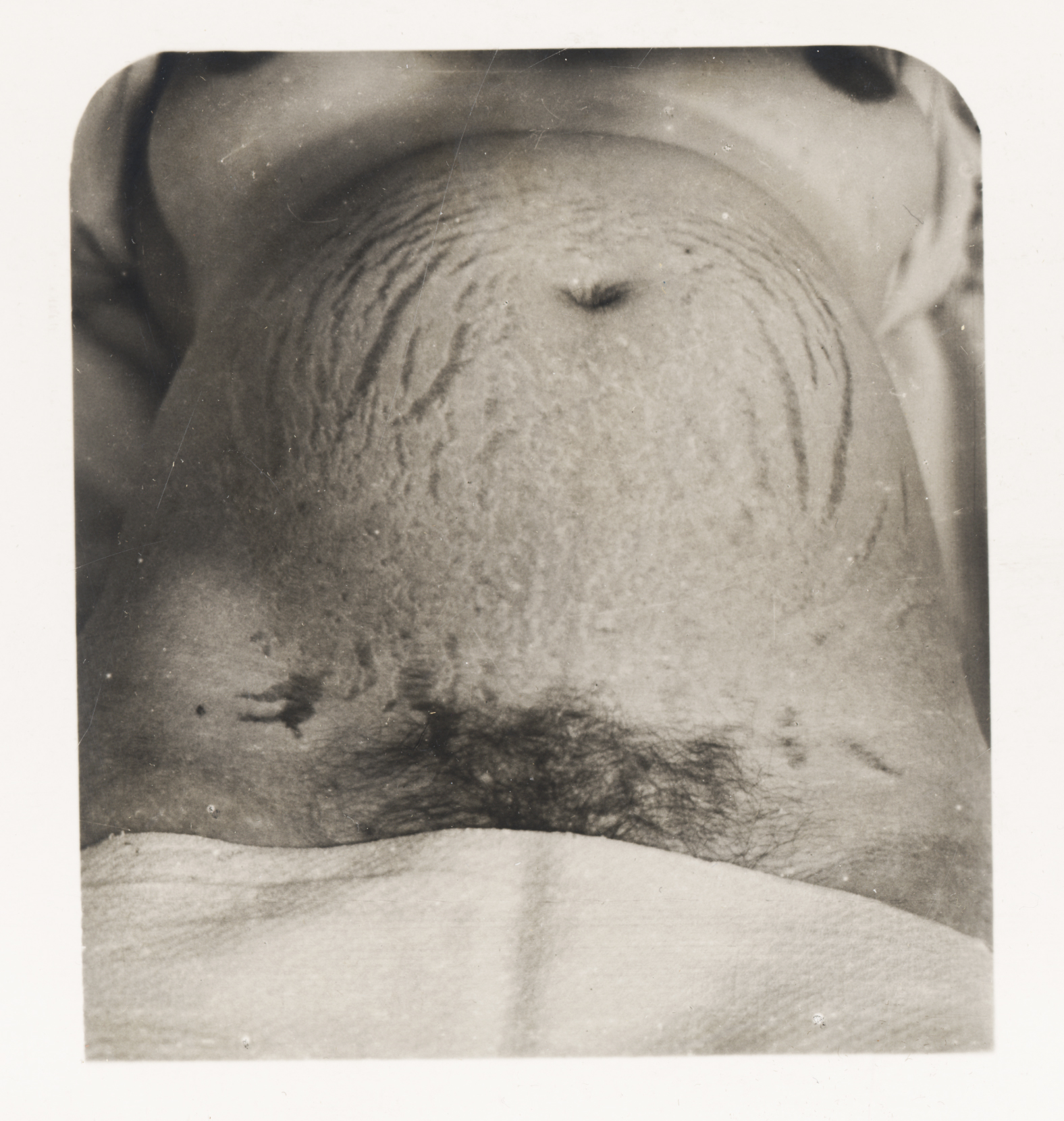
‘Abdomen of Gravid Multipara’ from The Edinburgh Stereoscopic Atlas of Obstetrics (published 190-) by G.F. Barbour Simpson
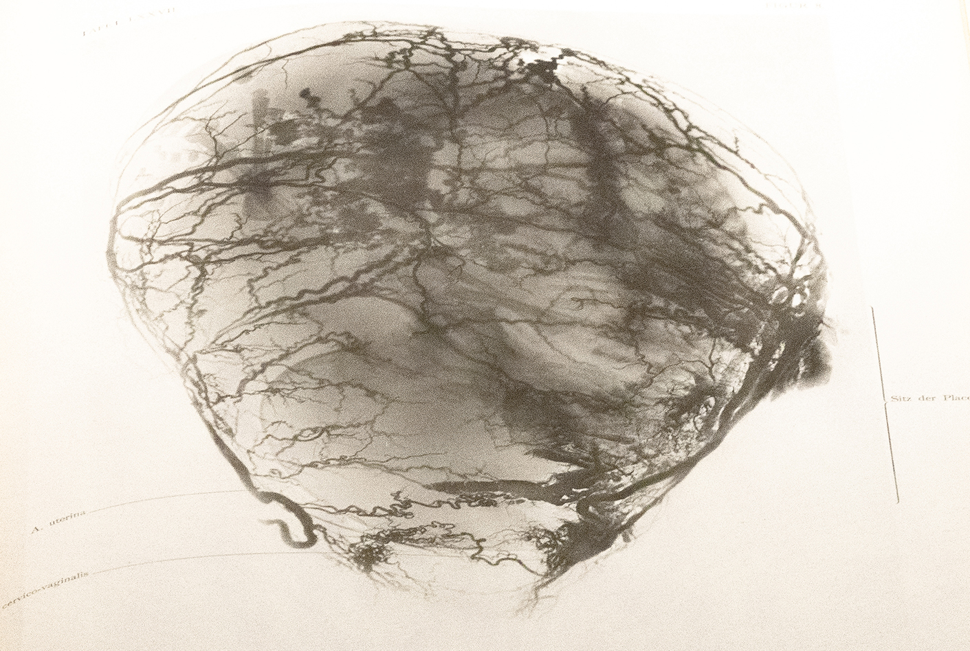
‘Placenta Praevia’ from The Edinburgh Stereoscopic Atlas of Obstetrics (published 190-) by G.F. Barbour Simpson

‘Left and Right Acromio-Posterior Position of the Shoulder’ from The Edinburgh Stereoscopic Atlas of Obstetrics (published 190-) by G.F. Barbour Simpson
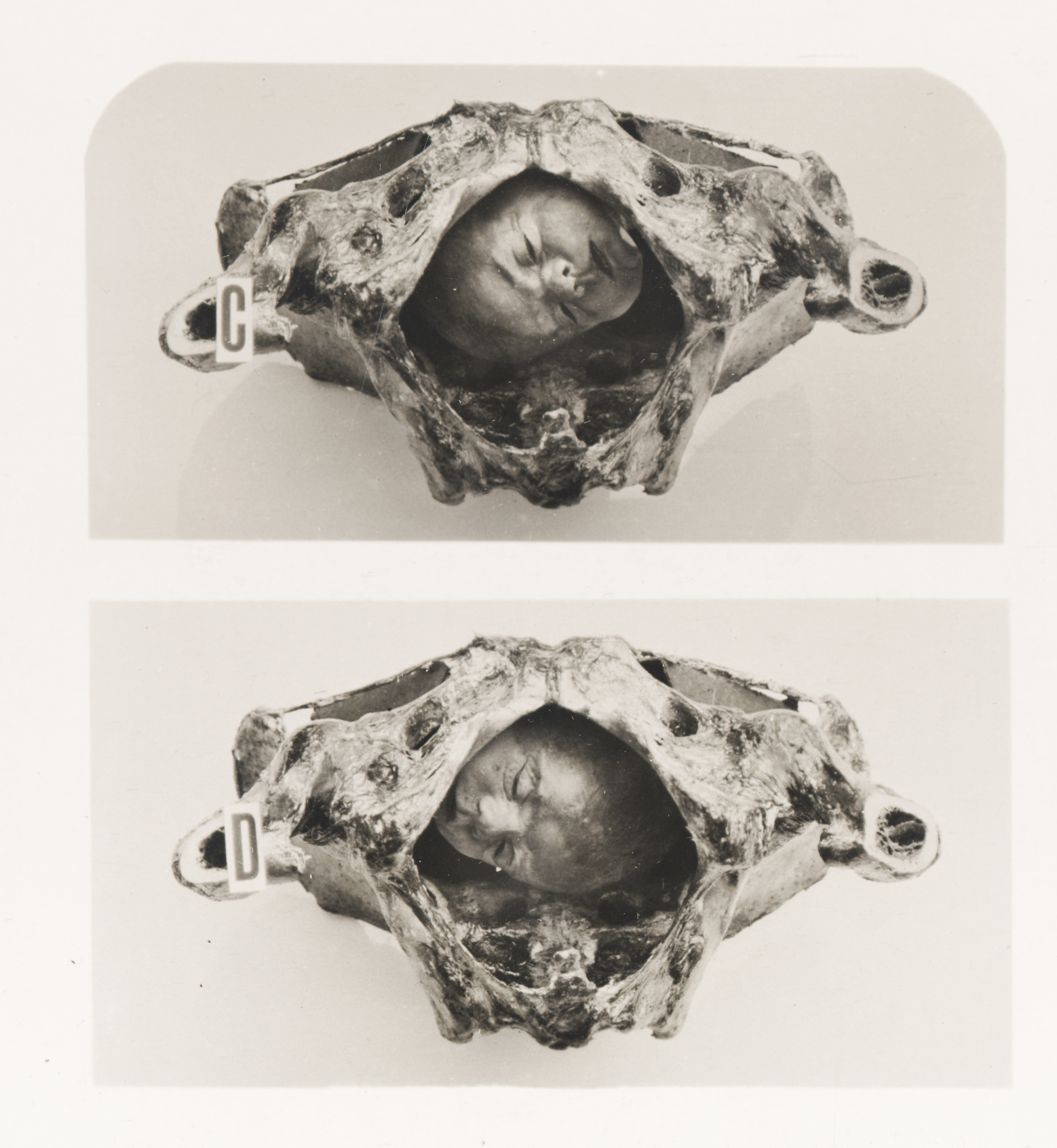
‘Left and Right Mento-Anterior Position of Face’ from The Edinburgh Stereoscopic Atlas of Obstetrics (published 190-) by G.F. Barbour Simpson
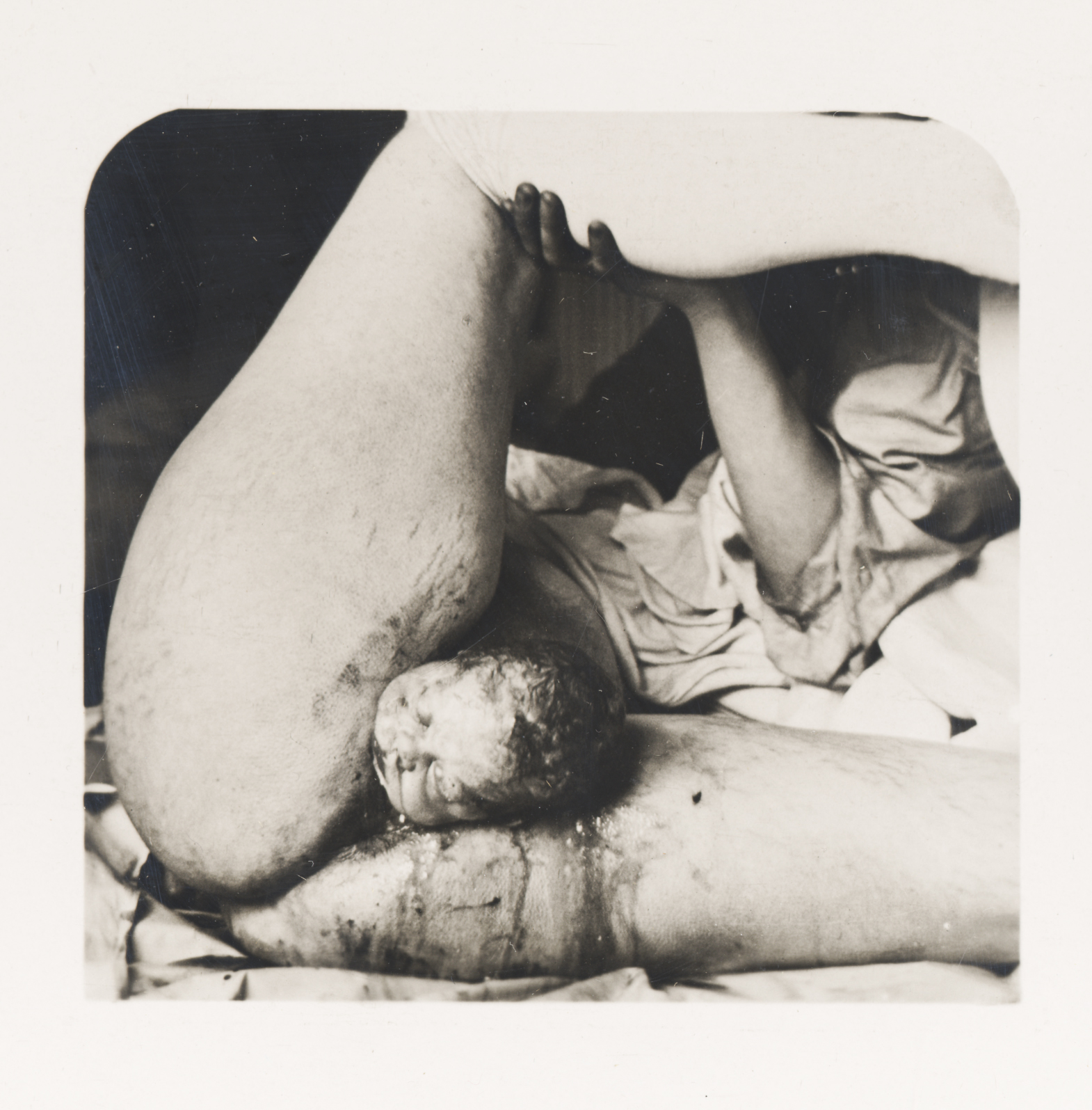
‘Normal Labor’ from The Edinburgh Stereoscopic Atlas of Obstetrics (published 190-) by G.F. Barbour Simpson

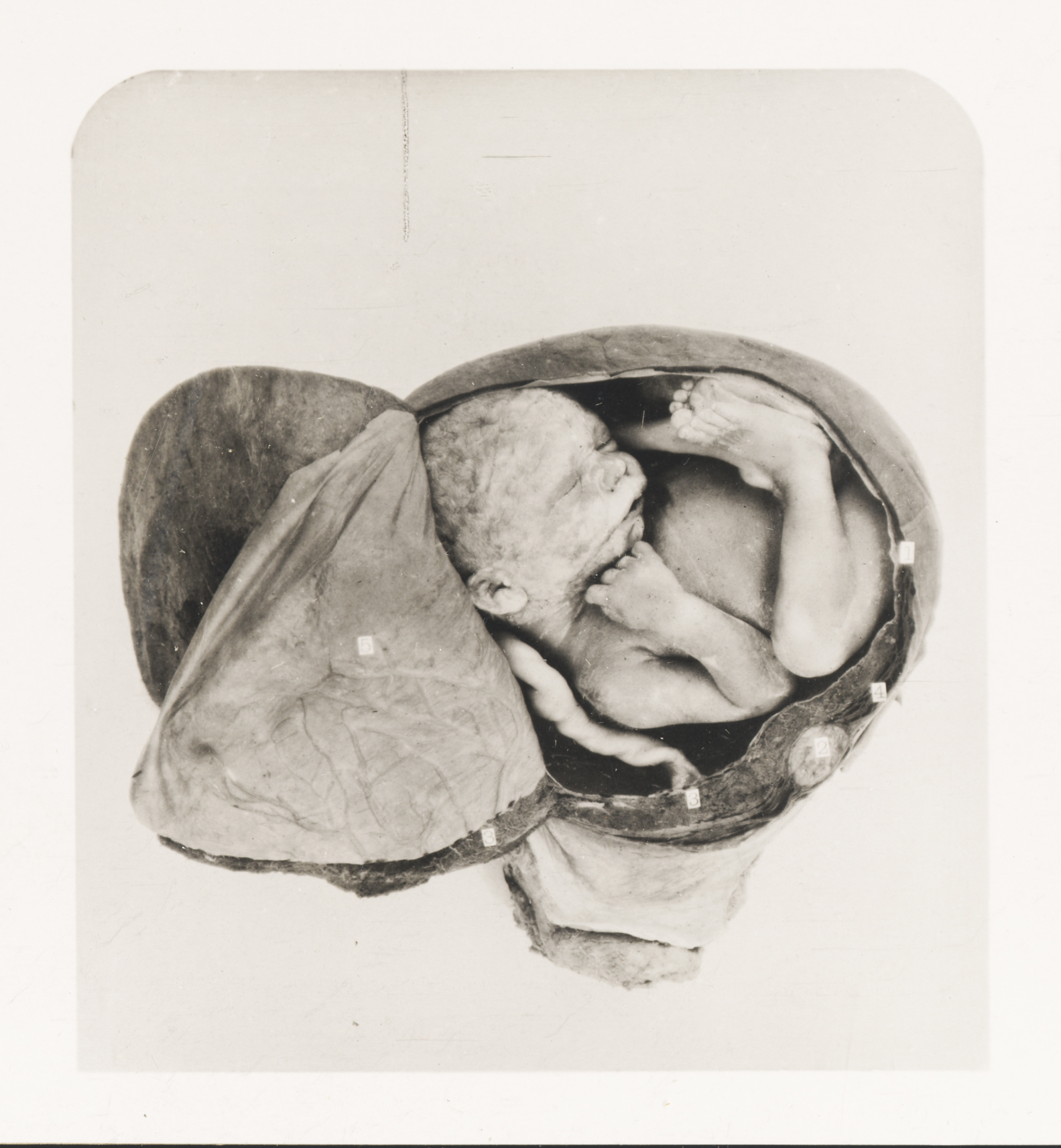
‘The Edinburgh stereoscopic atlas of obstetrics’ (published 190-) by G.F. Barbour Simpson, ed.
This is a photographic interpretation of the iconic image of the fetus in utero, whereby a full-term fetus is revealed by peeling back the uterus. Viewed through the stereoscope, the 3D presentation of the image expands the visual culture of the fetus in utero into a new dimension and serves as a precursor to the 3D ultrasound images that emerge decades later. Similar to other images in the Atlas, the image, when viewed through the stereoscope, is a haunting depiction of death brought into full relief.
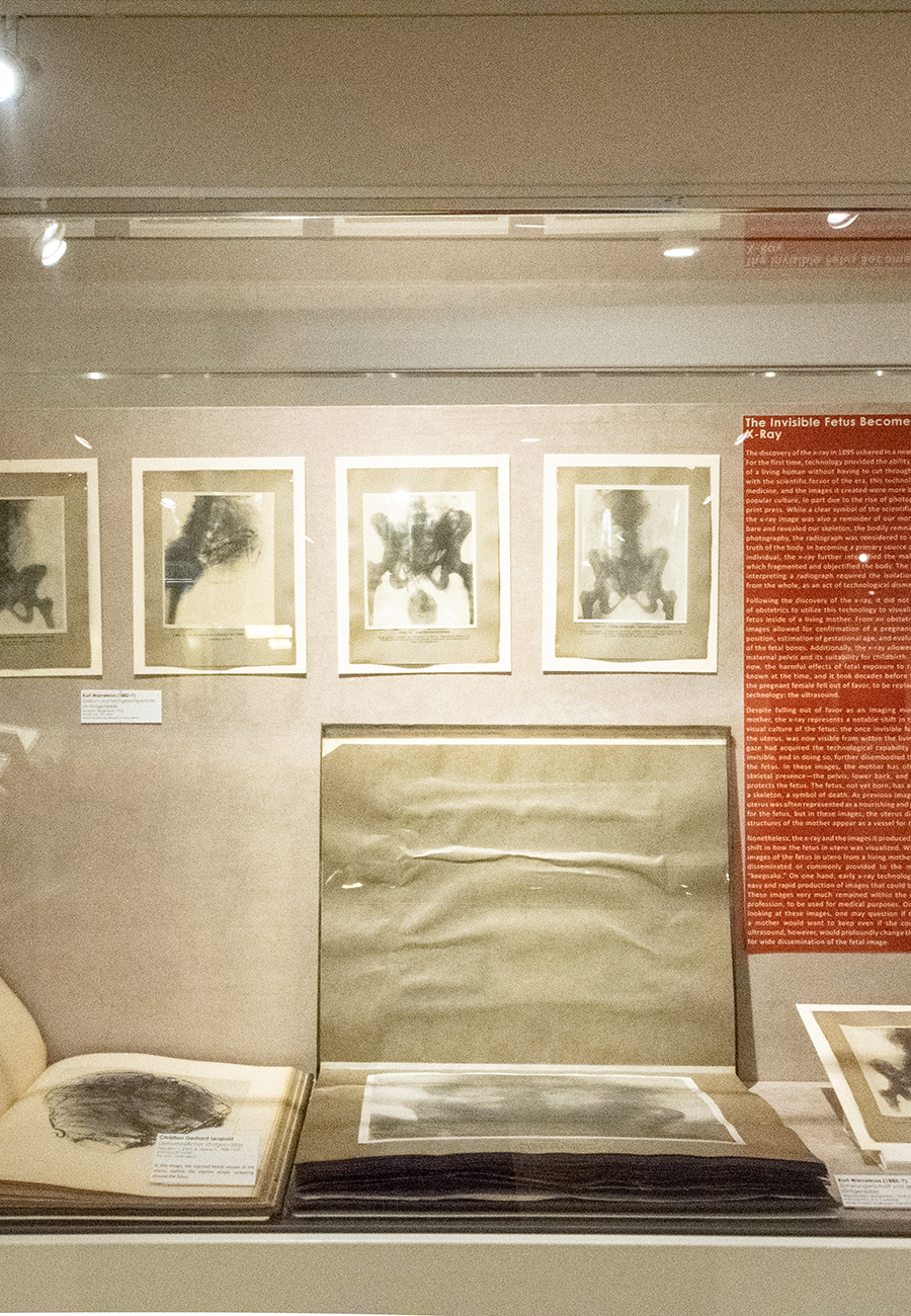
The Invisible Fetus Becomes Visible: The X-ray
The Invisible Fetus Becomes Visible: The X-Ray
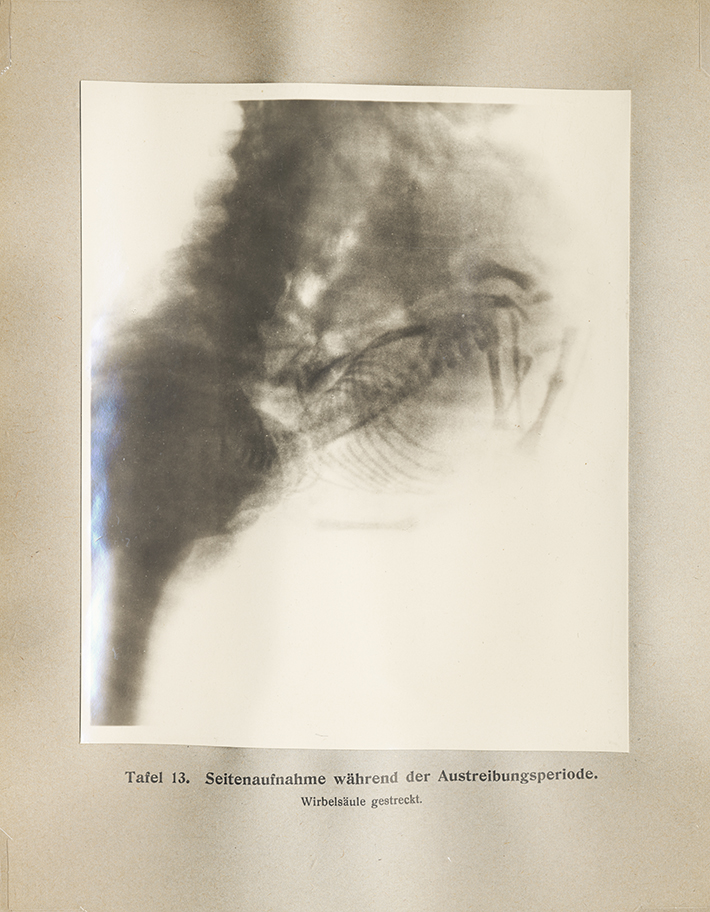
The discovery of the x-ray in 1895 ushered in a new era of medical imaging. For the first time, technology provided the ability to visualize the interior of a living human without having to cut through one’s skin. In keeping with the scientific fervor of the era, this technology was widely used in medicine, and the images it created were more broadly disseminated in popular culture, in part due to the rise of photography and the popular print press. While a clear symbol of the scientific progress of humanity, the x-ray image was also a reminder of our mortality, as it stripped us bare and revealed our skeleton, the bodily remnants of death. Similar to photography, the radiograph was considered to represent the objective truth of the body. In becoming a primary source of information about an individual, the x-ray further intensified the male-driven medical gaze, which fragmented and objectified the body. The process of creating and interpreting a radiograph required the isolation of parts of the body from the whole, as an act of technological dismemberment.
Following the discovery of the x-ray, it did not take long for the field of obstetrics to utilize this technology to visualize, for the first time, a fetus inside of a living mother. From an obstetrical perspective, these images allowed for confirmation of a pregnancy, assessment of fetal position, estimation of gestational age, and evaluation for abnormalities of the fetal bones. Additionally, the x-ray allowed for assessment of the maternal pelvis and its suitability for childbirth. While it seems obvious now, the harmful effects of fetal exposure to radiation were not well known at the time, and it took decades before the use of the x-ray on the pregnant female fell out of favor, to be replaced by a new, emerging technology: the ultrasound.
Despite falling out of favor as an imaging modality of the pregnant mother, the x-ray represents a notable shift in the development of the visual culture of the fetus: the once invisible fetus, still hidden within the uterus, was now visible from within the living mother. The medical gaze had acquired the technological capability to see what was once invisible, and in doing so, further disembodied the mother and isolated the fetus. In these images, the mother has often been reduced to a skeletal presence—the pelvis, lower back, and hips—that cradles and protects the fetus. The fetus, not yet born, has already been reduced to a skeleton, a symbol of death. As previous images attest, the maternal uterus was often represented as a nourishing and protective environment for the fetus, but in these images, the uterus disappears and the bony structures of the mother appear as a vessel for the fetal skeleton.
Nonetheless, the x-ray and the images it produced represents a significant shift in how the fetus in utero was visualized. While the x-ray produced images of the fetus in utero from a living mother, they were not widely disseminated or commonly provided to the mother as a pregnancy “keepsake.” On one hand, early x-ray technology did not facilitate the easy and rapid production of images that could be provided to mothers. These images very much remained within the domain of the medical profession, to be used for medical purposes. On the other hand, upon looking at these images, one may question if this was an image that a mother would want to keep even if she could. The emergence of ultrasound, however, would profoundly change these dynamics, allowing for wide dissemination of the fetal image.
The Invisible Fetus Becomes Visible: The X-Ray
‘Geburtshilflicher röntgen-atlas’ (published 1908-1909) by Christian Gerhard Leopold
In this image, the injected blood vessels of the uterus outline the uterine shape, wrapping around the fetus.
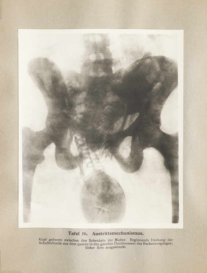
‘Geburtshilflicher röntgen-atlas’ (published 1908-1909) by Christian Gerhard Leopold
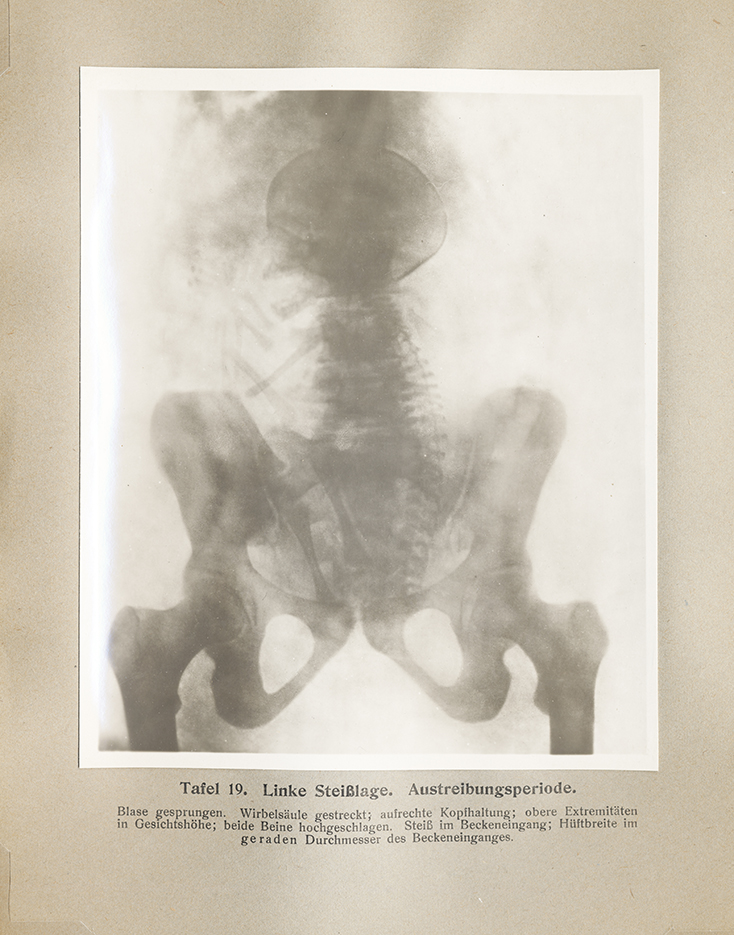
‘Geburtshilflicher röntgen-atlas’ (published 1908-1909) by Christian Gerhard Leopold
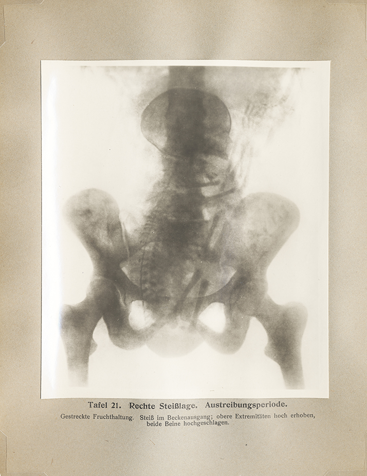
‘Geburtshilflicher röntgen-atlas’ (published 1908-1909) by Christian Gerhard Leopold
‘Geburtshilflicher röntgen-atlas’ (published 1908-1909) by Christian Gerhard Leopold
Twinning the Fetus in Utero
Twinning the Fetus in Utero
Although the phenomenon of twins and multiples is as old as human reproduction itself, their prominence in obstetrical textbooks suggests that they were medically prevalent if not also challenging, as well as culturally novel. The notion that twins develop a special, life-lasting bond in utero has a long history, and in the images presented here, they are occasionally depicted embracing or facing each other, as if deep in conversation. On other occasions, they appear like nesting dolls inside an egg or puppets attached by string-like umbilical cords.
Early modern practitioners differentiated between identical and fraternal twins, though the nature of both was inscribed in a larger debate about the origins of human conception. Were humans pre-formed at the moment of their conception, merely requiring time and energy to grow to adult size? And if so, did the preformed parts reside in the female egg (ovist theory) or the male sperm (spermist or animalculist theory)? If humans were not pre-formed, then were they the product of “epigenesis”—that is, a spontaneously generated lower order form who morphologizes into the more complex full-term fetus over the course of the gestation period?
More in-depth investigations of fetal biology in the nineteenth century would provide answers to these questions, just as anatomical investigations of the placement of twins in utero would reveal their precise configuration. While identical twins are the result of a single fertilized egg that grows in the same amniotic sac, fraternal twins emerge from separately fertilized eggs that develop in distinct sacs. The notion that an egg or ovum can split into two or more clones, or that multiple eggs can simultaneously be fertilized, was and remains a fascinating biological phenomenon that has been studied across the animal kingdom.
Competing theories aside, multiple fetuses presented practitioners with a particular set of challenges during childbirth. The medical concern over twins was focused on managing risk. This was especially the case in the early modern period before the advent of imaging technology that would reveal twins in utero prepartum, knowledge that would in the modern period allow practitioners to prepare for the various eventualities of delivery. In the absence of x-ray vision, such practitioners as the eighteenth-century French midwife Mme du Coudray (ca. 1712–94) warned that all fetal presentations during delivery should be treated with the possibility of twins or multiples in mind. For instance, one should not assume that an arm and a leg presenting themselves are necessarily from one child. Additionally, twins safely nestled in utero often appear oriented in the opposite direction, meaning that one of the two would present itself in the risky breech (foot-first) position.
The advent of the x-ray and then ultrasound facilitated delivery of twins and permitted practitioners to anticipate potential birthing complications and devise delivery strategies accordingly. In fact, twins are often by default categorized as high risk births and the medical establishment today views favorably on C-sections for such delivery, an operation that was considered very dangerous in the early modern period.
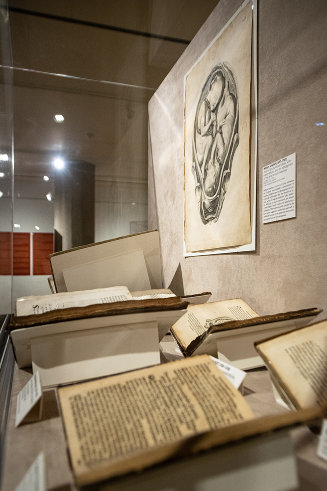
Twinning the Fetus in Utero
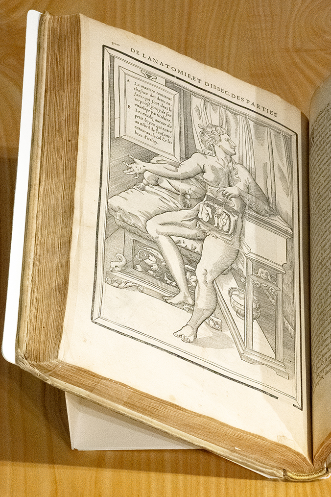
‘La dissection des parties du corps humain’ (published 1546) by Charles Estienne (1504–ca. 1564)
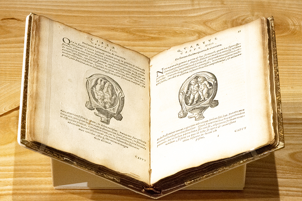
‘De conceptu, et generatione hominis’ (published 1587) by Jakob Rüff (1500–1558)
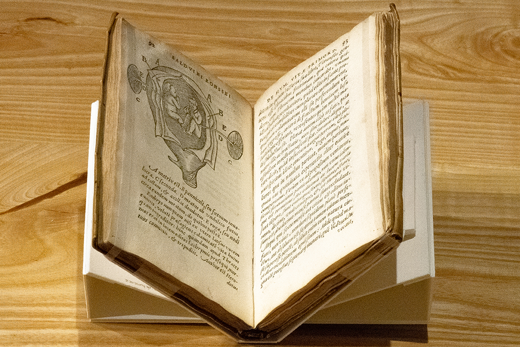
‘De humanæ vitæ primordiis’ (published 1594) by Baudouin van Ronss (?–1596)
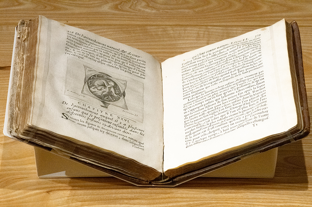
‘Des maladies des femmes grosses et accouchees’ (published 1668) by Jakob Rüff (1500–1558)
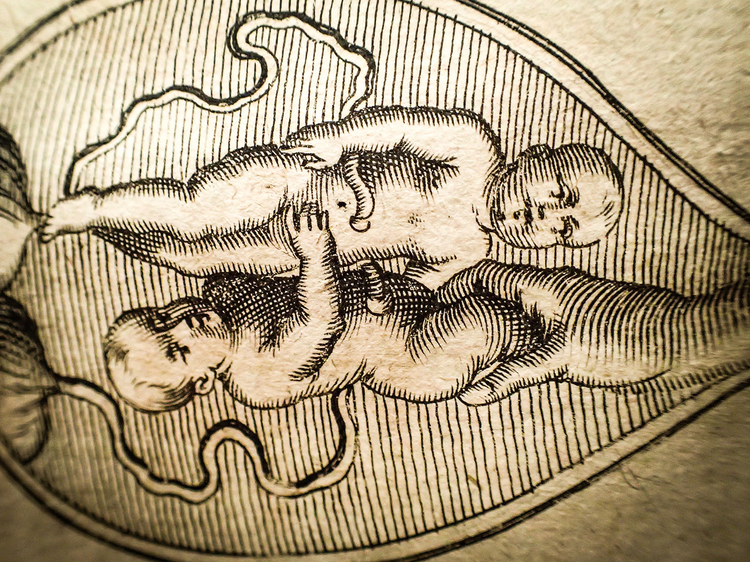
‘Anmerckungen von der weiblichen Geburt’ (published 1676) by Cosme Viardel (fl. 1671–1674)
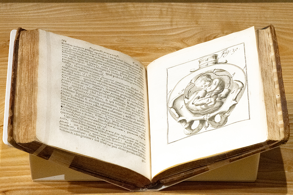
‘Operationes chirurgicae novum lumen exhibentes obstetricantibus’ (published 1701) by Hendrik van Deventer (1651–1724)
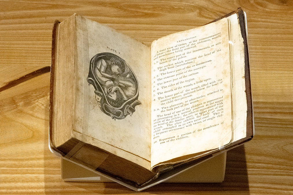
‘The midwife’s practical directory’ (published 1836) by Thomas Hersey
Charting Fetal Development: from Generation to Reproduction
Charting Fetal Development: from Generation to Reproduction
Before about 1800, the term used for conception by laymen and medical experts alike was “generation.” This was eminently suitable given the prevailing Christian worldview and its etymological kinship with the biblical concept of creation or “genesis.” From about 1800 onward, however, “reproduction” became the preferred term. Its underlying notion of copying can be understood against the backdrop of rapid industrialization and the introduction of mechanical means of mimicking human labor that extended from the factory floor to new technologies of imaging, such as the advent of lithography and photography.
Up until about 1800, there were also a variety of competing theories for the origins of human life, though the argument for divine design was generally accepted as the starting point for any particular theory in the Christian West. The two most popular theories of human origins debated in the early modern period were preformationism and epigenesis.
According to preformationism, humans exist in their complete form even before they are conceived, requiring merely time and nourishment as embryos in order to grow from miniature to adult size. The question that remained was: where did preformed life reside before conception—in the female ovum or the male sperm? According to the ovist model, life originated in the female egg or ovum. Adherents of the spermist (or animalculist) theory believed, on the other hand, that the preformed parts were found in the head of the male sperm, based on their observation of sperm motility in semen samples examined with the newly invented microscope. The theory of epigenesis, on the other hand, held that humans developed spontaneously from an undifferentiated mass or lower order being to a more complex one over a series of morphological transformations resulting in a full-term infant at nine months.
Both theories could accommodate Christian theology given that the origins of both ‘mini me’ (preformationism) and ‘the blob’ (epigenesis) was a purposive God. Fetal development in the early modern works featured here showcase this range of conception theories. The fetus comes under many guises, from a nondescript mass to a recognizable baby, as well as everything in between: skeletons, doughboys, and little people.
Of these two early modern theories of conception, epigenesis more clearly aligned with nineteenth-century breakthroughs in evolutionary biology, which provided a new way of looking at and visually depicting the fetus in utero. In the grand scheme of the evolution of living things from a single common ancestor over deep time, fetal development was a rapid, singular process. Briefly put, evolutionary biologists—inspired by the English naturalist Charles Darwin’s ground-breaking work On the Origin of Species (1859)—understood fetal transformations as a developmental series. The fetus morphologized during its gestation period in utero, and in this process, became more complex. A variety of biologists, including German theorists from Samuel Thomas von Sömmering (1755–1830) to Ernst Haeckel (1834–1919), contributed to the creation of a new graphic convention of presenting the fetus’ serialized development in utero. By often removing the fetus from the uterus and presenting its developmental narrative in sequence, the fetus attained autonomy and a sense of personhood.
The serial or sequential model of human development also permitted biologists to distinguish more clearly between the embryo and the fetus, each of which aligned with specific periods of human gestation. This demarcation, in turn, has fueled a variety of debates over fetal personhood in modern-day litigation, from the permissibility of late-term abortion to determining parentage in the cases of in vitro fertilization.
Charting Fetal Development: from Generation to Reproduction
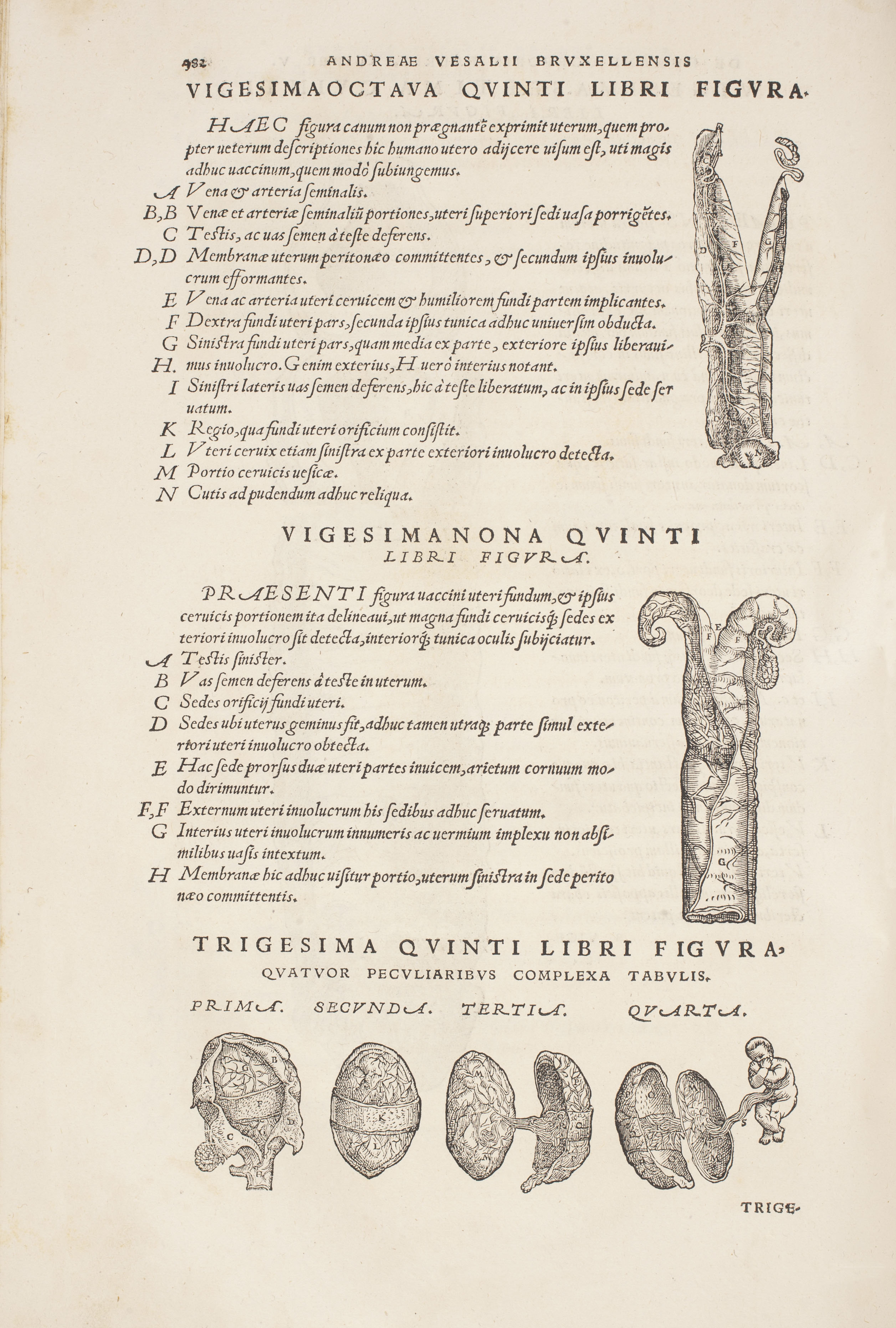
‘Scholae medicorum Patauinae professoris de humani corporis fabrica libri septem’ (published 1543) by Andreas Vesalius (1514–1564)

‘The history of generation’ (published 1651) by Nathaniel Highmore (1613–1685)
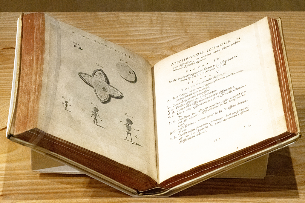
‘Spicilegivm anatomicvm, continens observationum anatomicarum rariorum centuriam unam’ (published 1670) by Theodor Kerckring (1640–1693)
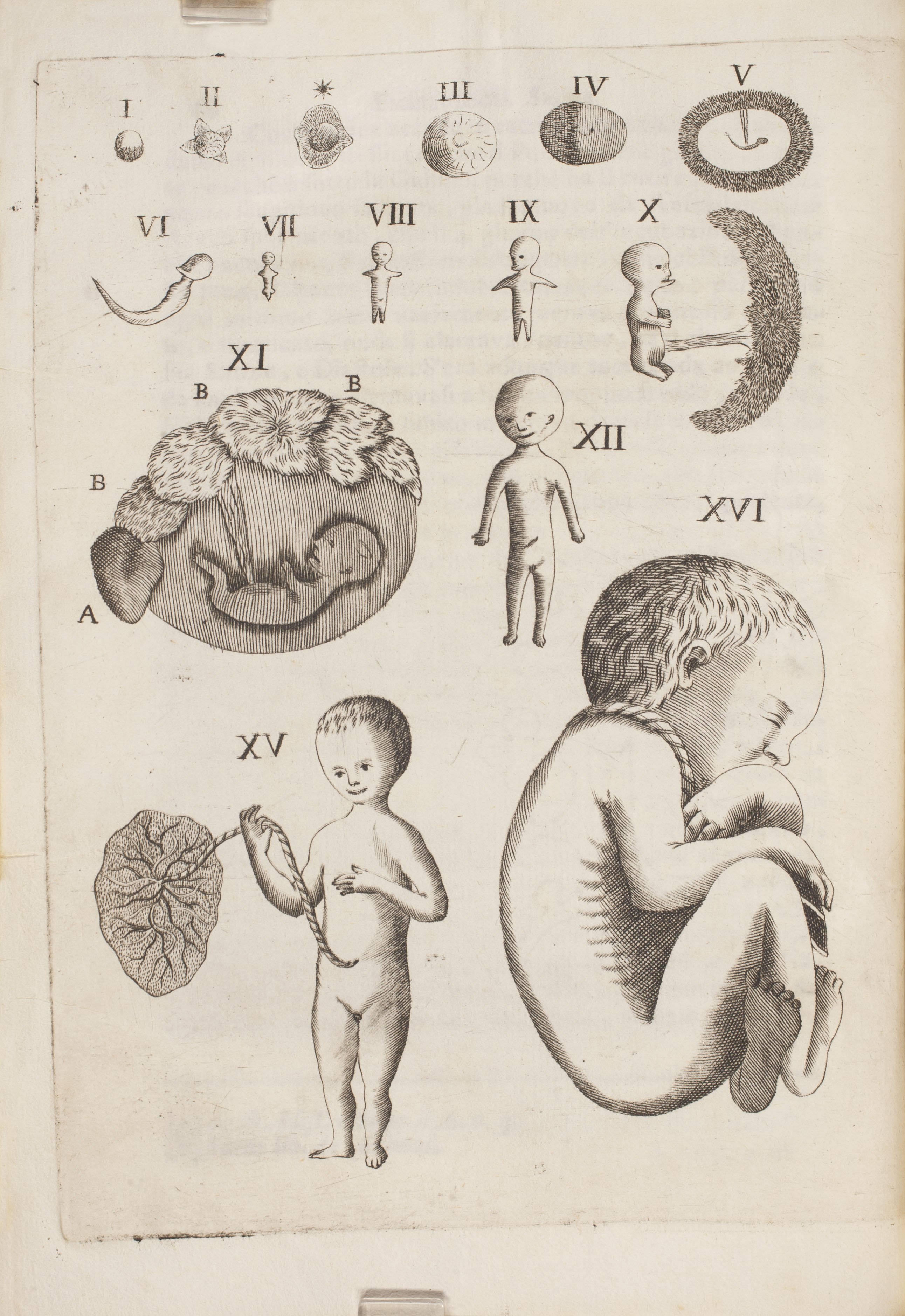
‘Embriologia sacra’ (published 1745) by Francesco Emmanuele Cangiamila (1702–1763)
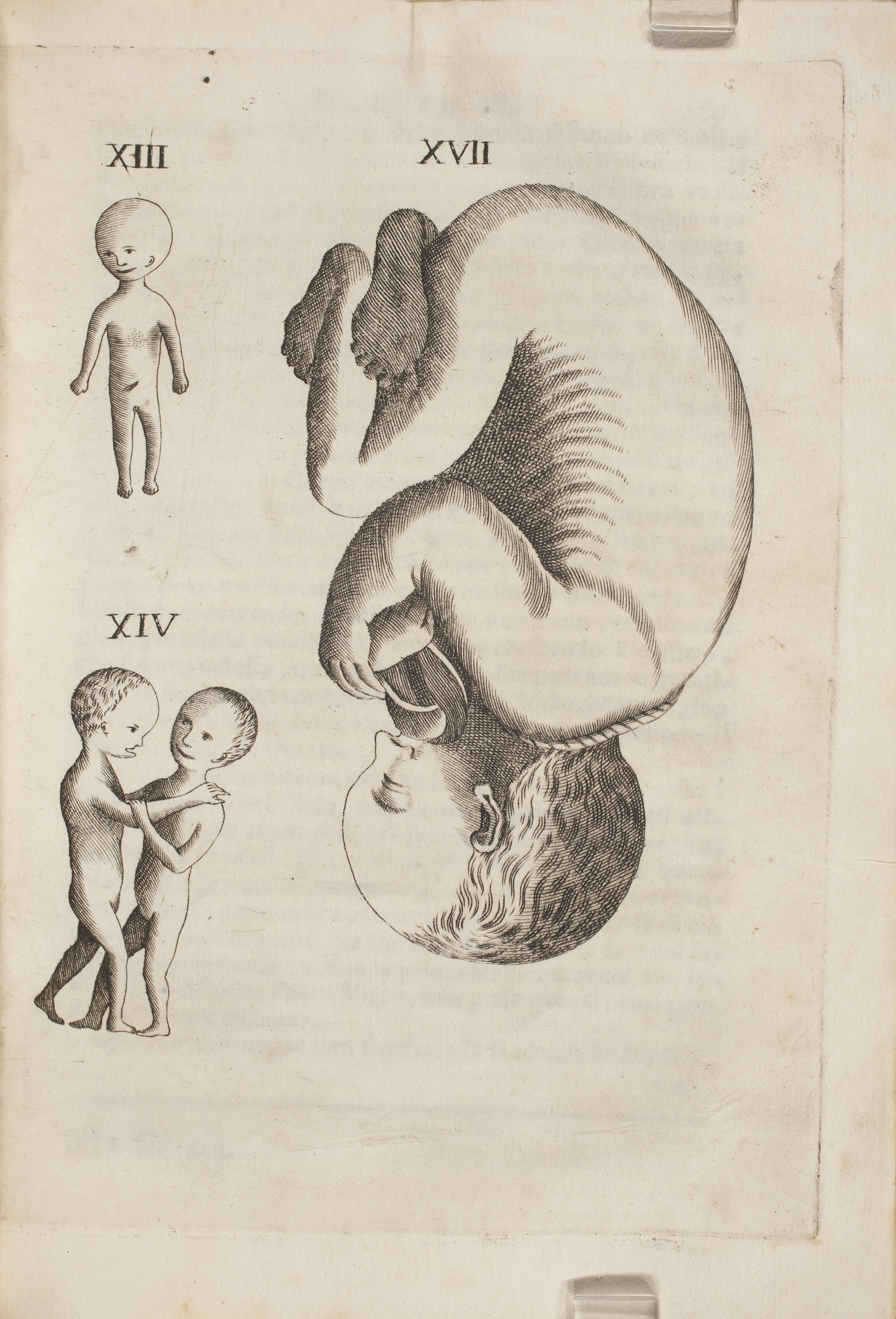
‘Embriologia sacra’ (published 1745) by Francesco Emmanuele Cangiamila (1702–1763)
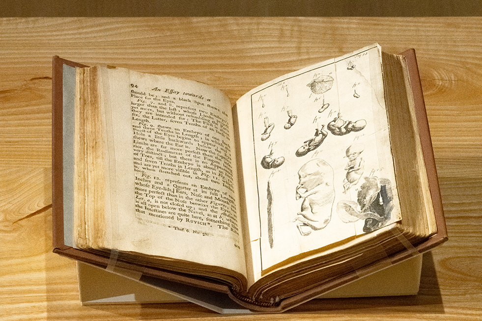
‘An essay towards a complete new system of midwifery’ (published 1751) by John Burton (1710–1771)
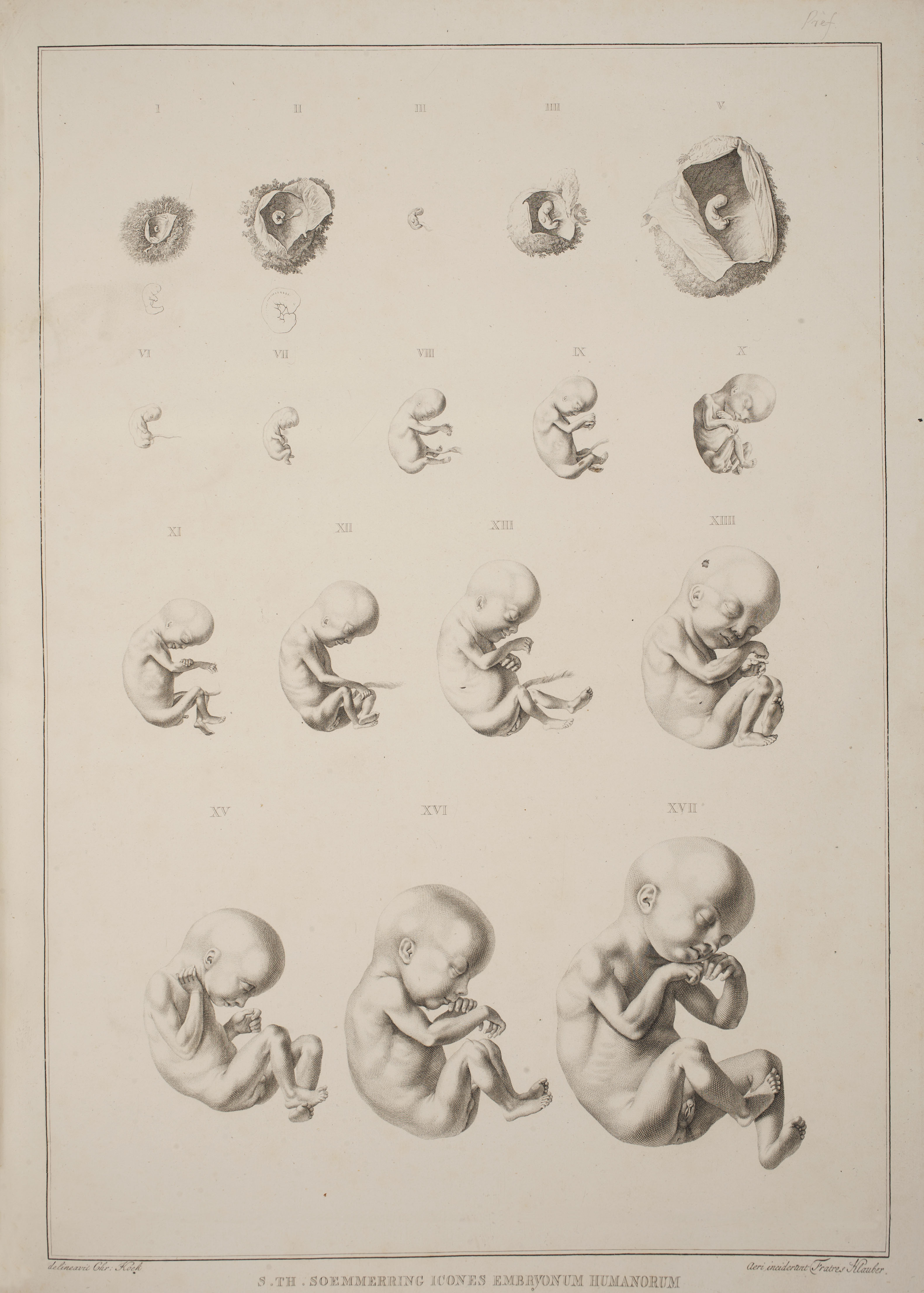
‘Icones embryonum humanorum’ (published 1799) by Samuel Thomas von Soemmerring (1755–1830)
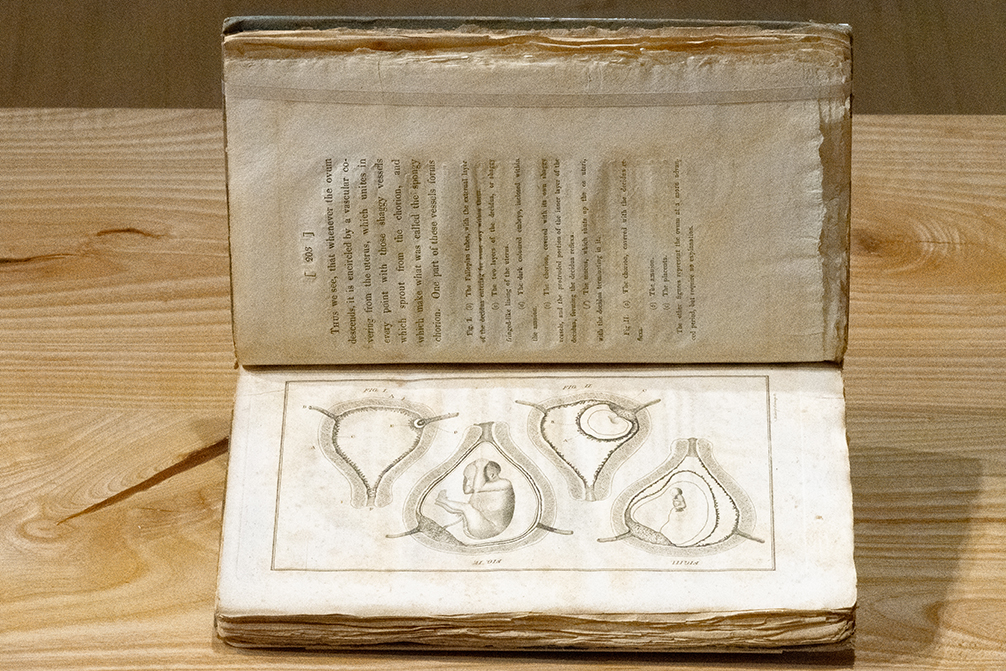
‘The anatomy of the gravid uterus’ (published 1808) by John Burns (1774–1850)

‘Die gebärmutter und das ei des menschen in den ersten schwangerschaftsmonaten’ (published 1832) by Burkhard Eilhelm Seiler (1779–1843)
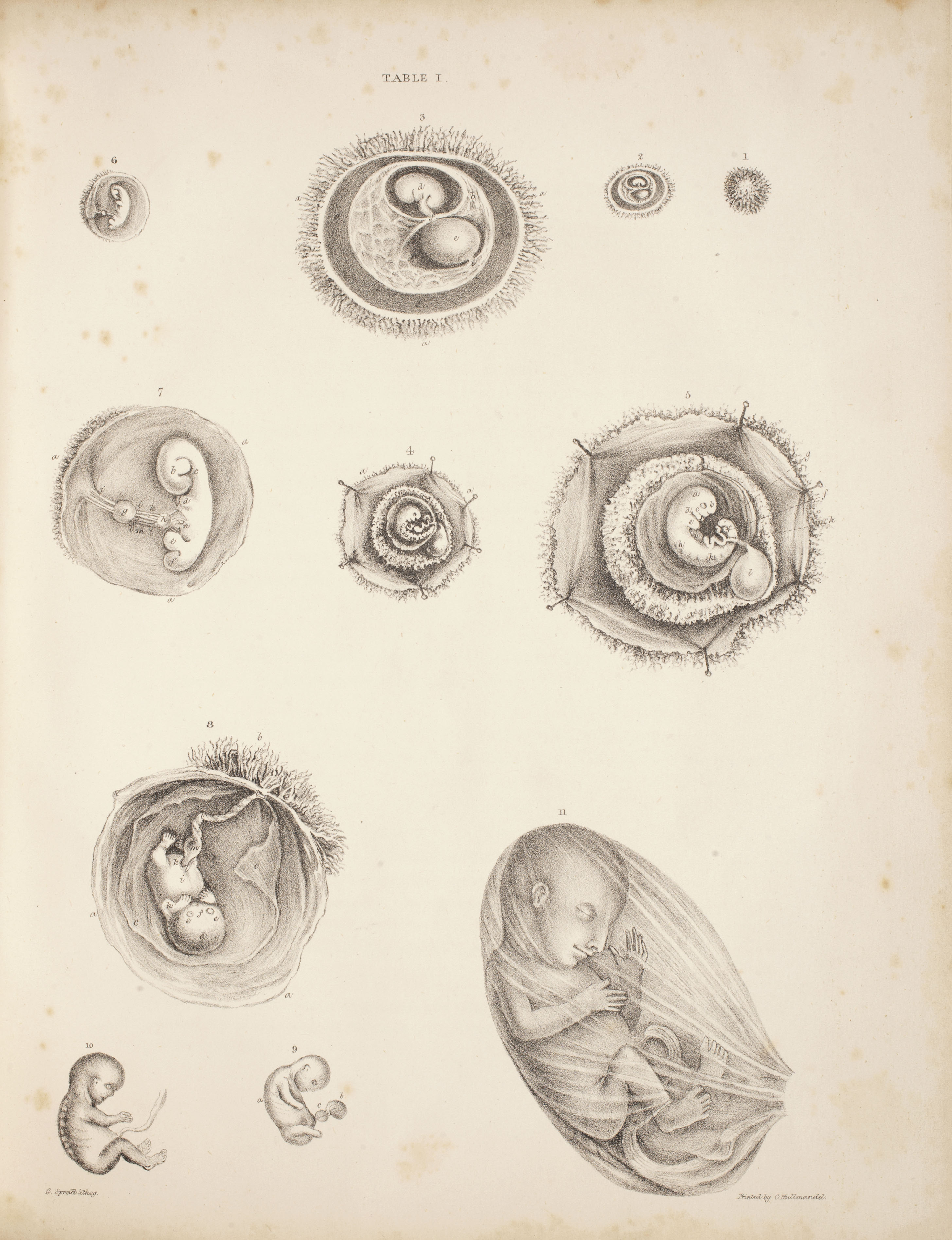
‘Supplement to obstetric tables’ (published 1835) by G. (George) Spratt
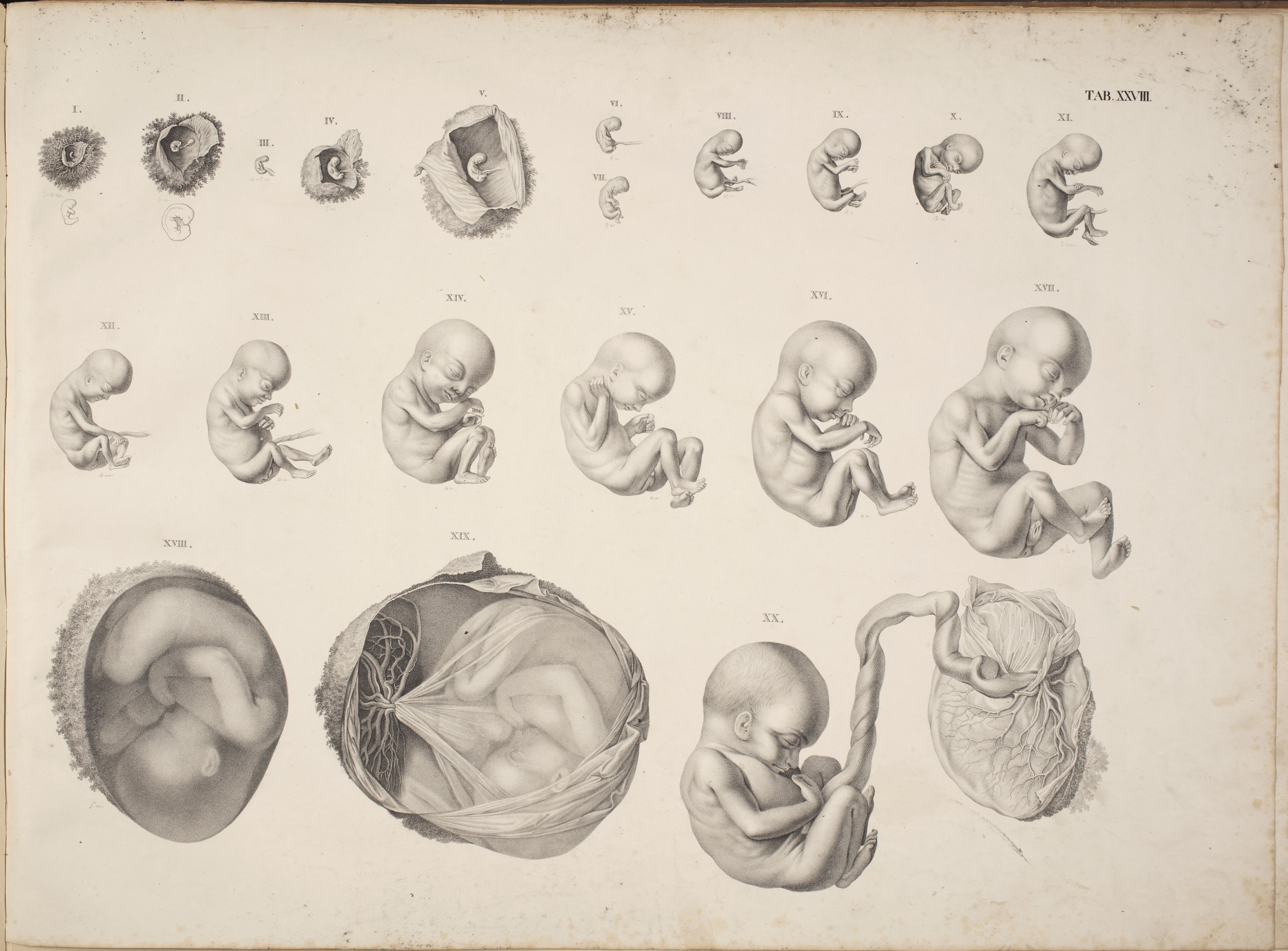
‘Geburtshülflicher atlas in 48 tafeln und erklärendem texte’ (published 1835-44) by Hermann Friedrich Kilian (1800–1863)

‘Natürliche schöpfungsgeschichte, gemeinverständliche wissenschaftliche vorträge’ (published 1870) by Ernst Haeckel (1834–1919)
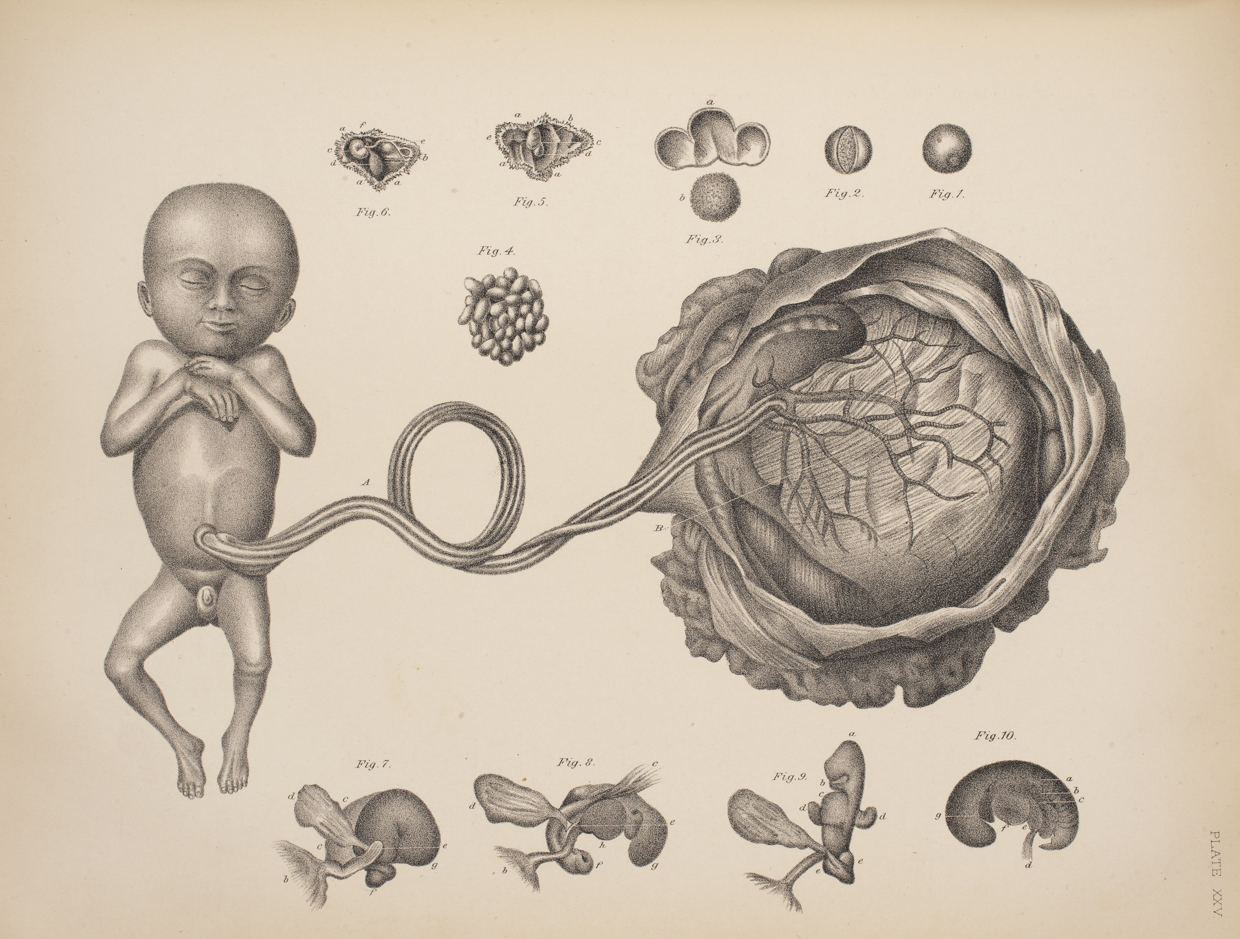
‘An illustrated encyclopaedia of the science and practice of obstetrics’ (published 1890) by F. H. Getchell, ed.
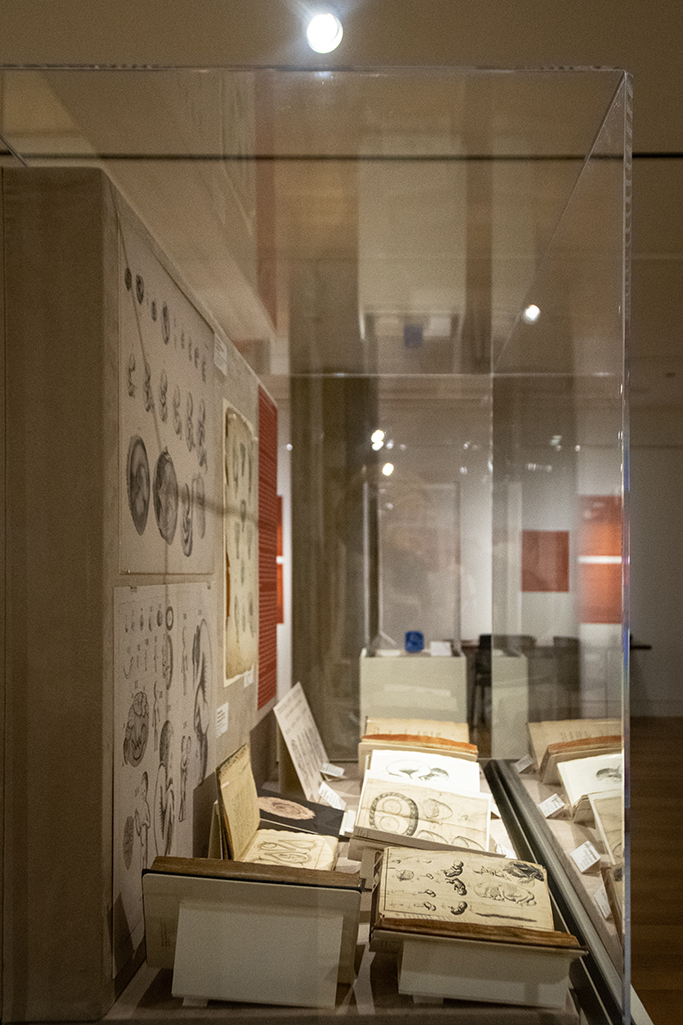
Physician’s Anatomical Aid
Physicians’ Anatomical Aid
Unlocking Victorian Anatomy: Similar to other flap anatomy images presented throughout the exhibition, this educational tool presents serialized fetal development within the disembodied maternal body. Interestingly, the physical format of this anatomical aid adhered to Victorian concerns of voyeurism and modesty when it comes to the female body and pregnancy. The images of both the male and female genitalia, including the gravid uterus, were separated from other parts of the body and literally kept behind a lock and key in a separate compartment of this work, presumably to prevent improper and voyeuristic viewing. Locked away for decades, these hidden images were only revealed when the locked flap was cut to allow access to them for display in this exhibit. Viewer discretion playfully advised.

‘Physicians’ Anatomical Aid’ (published 1888)
The Static Maternal Vessel: Through a series of cross sections, this source presents active fetal development, embodied male anatomy, and a static maternal vessel. In the half illustrating female anatomy, the woman’s thighs are severed, revealing bone and flesh. Although the fetus is presented as alive and developing inside her, the mother’s amputated legs suggests she is deceased. This contrast between the presumably dead mother and the growing fetus alienates her from her organs of generation while removing any notion of agency over her body and pregnancy. The comparative wholeness of the fetus simultaneously emphasizes its personhood and maternal disembodiment. By contrast, the cross sections of the male in this work are alive: his legs are intact, not severed, and extend off the page, while his penis is portrayed as both flaccid and erect. While the man and the fetus are both represented as active subjects, the woman is depicted as a static vessel. Caption written by Anya Dunaif
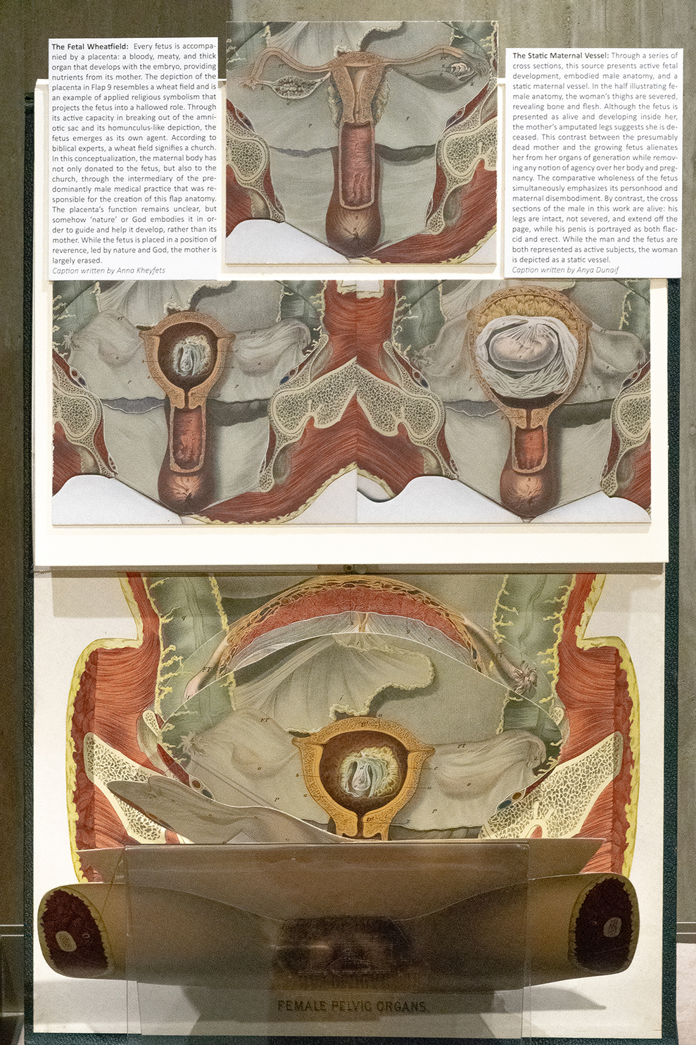
‘Physicians’ Anatomical Aid’ (published 1888)
The Fetal Wheatfield: Every fetus is accompanied by a placenta: a bloody, meaty, and thick organ that develops with the embryo, providing nutrients from its mother. The depiction of the placenta in Flap 9 resembles a wheat field and is an example of applied religious symbolism that projects the fetus into a hallowed role. Through its active capacity in breaking out of the amniotic sac and its homunculus-like depiction, the fetus emerges as its own agent. According to biblical experts, a wheat field signifies a church. In this conceptualization, the maternal body has not only donated to the fetus, but also to the church, through the intermediary of the predominantly male medical practice that was responsible for the creation of this flap anatomy. The placenta’s function remains unclear, but somehow ‘nature’ or God embodies it in order to guide and help it develop, rather than its mother. While the fetus is placed in a position of reverence, led by nature and God, the mother is largely erased. Caption written by Anna Kheyfets
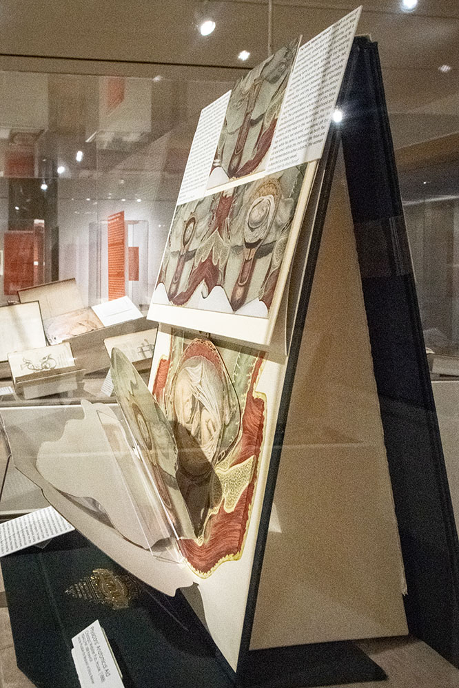
The Fetal Image Goes Public
Ultrasound and the Fetal Image
The development of the ultrasound as an imaging modality and its subsequent use in obstetrics marks a pivotal period in the visual culture of the fetus in utero. Arguably the most recognizable medical image, the fetal sonogram has become iconic. Over the course of its technological development, the ultrasound has attained increasing capability to visualize in greater detail and complexity the fetus in utero.
Occurring within the context of the increased medicalization and socialization of pregnancy, the fetal ultrasound has become a routine clinical procedure during pregnancy, while the images it produces have contributed to a public narrative around pregnancy. This allowed the fetal sonogram, in combination with sensationalized photography and the rise of the internet and social media, to project images of the fetus in utero into the public domain, to be curated in cyberspace by the parent(s)-to-be and deployed as a political tool.
But why the fetal ultrasound? The emergence and dissemination of the sonogram as the dominant image within the visual culture of the fetus can be traced back to the technology itself and how it came to be utilized in obstetrical practice. Compared to other imaging modalities (CT, MRI, X-ray), the ultrasound is relatively easier to use, safer (avoids radiation exposure), less expensive, and importantly, produces moving images that can be viewed in real-time. For these reasons, ultrasound has become a versatile and commonly used technology in medicine, including in obstetrics. From a clinical perspective, the ultrasound provides medically relevant information about both the fetus and pregnant woman, including confirmation of pregnancy, fetal measurements, fetal anatomy including abnormalities, location of the placenta, and amount of amniotic fluid. Despite its clinical relevance, the ubiquitous use of ultrasound in obstetrics has been criticized for contributing to the medicalization of pregnancy. The images it produces, on the other hand, have been critiqued for contributing to the emergence of the public pregnancy.
However, the fetal sonogram did not attain iconic status until the images achieved enough clarity that they could be easily interpreted and, in turn, readily distributed to parents-to-be for further distribution beyond the clinic and into the public domain. Fetal sonograms from the early era of ultrasound technology are unrecognizable when compared to contemporary images. A brief timeline of the obstetric ultrasound illustrates how the visual culture of the fetal sonogram evolved with the technology to its present-day aesthetic and how, in the process, the sonogram became socially “readable” through its presentation of a discernible fetus in utero.
An early ultrasound from a 1966 publication measures the diameter of the fetal head, here referenced as two sharp peaks occurring 90mm apart. This A-mode (amplitude) technology was rudimentary and limited in its clinical utility.
This ultrasound image from a 1972 publication shows the diameter of the fetal head in cross-section. The B-mode (brightness) ultrasound technology used to produce these images marked a significant advancement in ultrasound imaging, allowing for the first two-dimensional images. While the early images from B-mode technology were black and white, by the mid-1970s, images could be rendered in gray scale and permitted greater distinction between bones, tissue, and fluid. This technological capacity is what permitted the construction and normalization of the now-common ultrasound imagery presenting the fetus as a cross-sectional ‘silhouette.’
A pivotal point in the development of the ultrasound was the introduction of real-time sonography in the late 1970s. Prior to this development, images were made in similar ways to taking a photographic snap-shot. For the first time, real-time ultrasound allowed the pregnant woman to directly participate in viewing the images as they are being created. This ability ushered in an era of patient-directed ultrasound, whereby the ultrasound experience extended beyond a medical procedure and took on broader social significance. The ultrasound industry participated in this transition by creating machines with screens that swivel towards the patient for easier viewing, with built-in printers to print “keepsake” images for the parents-to-be, and installation of imaging programs that enhance the images, with colored filters, for example.
This series of sonograms depicting the fetal body shows the incremental improvement of image clarity and reduction in background “noise” from 1985, 1990, and 1995 respectively, an era in which the obstetric ultrasound became common clinical practice during pregnancy.
The subsequent development of 3D rendering provided increasing clarity and life-like images that further enhanced the iconography of the fetal ultrasound. 4D technology has added motion to 3D images. The data from these ultrasound images is also being used to create 3D printed models of the fetus in utero.
The Fetus’ Very Public Debut: Life Magazine
The fetus’ very public debut in April 1965 as a photograph of an 18-week-old free-floating figure on the cover of Life magazine marks a pivotal point in the history of its visual culture. Taken by the photojournalist Lennart Nilsson (1922–2017), the image quickly gained iconic status after its publication, selling eight million copies within four days of its release. The magazine claimed that the image was the “first published photograph of a living fetus inside a uterus.” The accompanying photo essay is entitled “The Drama of Life Before Birth,” suggesting that the fetus is a character in a sweeping, propulsive visual narrative.
This photograph was one of a series of career-making images by Nilsson, who photographed fetuses like these in the context of ongoing abortion debates in his native Sweden. Although later in life he appeared agnostic on the issue of abortion, at the time this and other images were published in his book A Child is Born (1965), Nilsson was enthralled by the anti-abortion physician, Per Wetterdal. It was through Wetterdal that Nilsson gained access to the first fetuses he ever photographed at a Stockholm Women’s clinic.
While these images proved pivotal in debating new abortion legislation in Sweden at the time, they were presented in America less controversially, as examples of the wonders of the human biological world. What very few if any consumers of these iconic fetal images knew then, as now, was the degree of curation required to produce such healthy looking specimens: Nilsson used backlighting and other photographic techniques to present the fetus as if alive, where in fact he primarily photographed aborted fetuses. Feminist scholars have pointed out that although the cover image was of a living 18-week-embryo, taken using a wide angle lens and a surgical scope equipped with a flash beam, the other images in the spread were of embryos that had been surgically removed from the uterus and were no longer viable. In her work on the history of fetal and embryo specimen collection in the United States, Lynn Morgan writes of the irony—not just in Life, but in many other popular and scientific works on pregnancy and human development—of using images of dead fetuses to (mis)represent life. Morgan’s work reminds us that fetal images are open not only to interpretation, but also to manipulation.
Nilsson’s Life cover photo is exceptional not only because of its claim to be the first of a living embryo, but also because the placenta is visible. In recent decades, feminist scholars have interrogated the ways in which contemporary images of the fetus tend to erase the pregnant body, so that the fetus appears to float in isolation, what Rosalind Petchesky has called “a tiny man, or homunculus,” suspended in the empty vessel of the womb. Others have compared this trope to that of an astronaut free floating in the darkness of space, which Petchesky suggests speaks to an older, Hobbesian view of humans as “disconnected solitary, individuals.” In her work on abortion rhetoric, Celeste Condit argues that anti-abortion groups have obscured parts of the fetal image that might be perceived by the public as unpalatable or even monstrous, such as a reptilian “tail,” and instead have focused on more familiar images of small feet and hands. Condit argues that this manipulative technique leads us to see images of small feet or hands and visualize a fully-formed human in their place. Scholars argue that obscuring the maternal presence, or portraying pregnant bodies as empty vessels, promotes fetal “personhood” at the expense of the pregnant woman, who becomes invisible as the fetus becomes hyper-visible and fully human, purportedly but dubiously capable of attaining legal and political status.
The 1965 Life article was followed up twenty-five years later with a similar article, titled “How life begins,” again featuring Nilsson’s photography on the cover and in the article. The images in this issue demonstrate the new technological capabilities of photography, serving to consolidate the recognizability of fetal imagery in the popular press.
Viewers are encouraged to look at a reproduction of the full 1965 article at the interactive desk.
Written with Chloe Hall
The Social and Cultural Fetus
Although ultrasound remains a standard component of prenatal care, the fetal image has become what anthropologist Julie Roberts calls “hypermobile,” moving far beyond the realm of the clinic and clinical practice. Images of fetuses have become icons, almost instantly recognizable in social, commercial, and political contexts. And, like religious icons, seemingly untouchable images that serve evangelizing agendas. Examining modern and contemporary uses and interpretations of the image of the fetus in utero reveals shifting and contested attitudes about fetal “personhood,” the pregnant body, and the family unit.
Fetal sonograms are unique among medical images in the way they bridge private and public worlds. Medically, sonograms are performed in order to identify fetal anomalies, and may shape patients’ private decisions to continue with or to terminate a pregnancy. Yet, as more commonly deployed, fetal images are widely disseminated in the public sphere. For example, they may be used by happy parents-to-be to spread the news of their pregnancy or to “gender reveal” the fetus to their social networks. They are also used to advocate the idea that fetuses are “persons,” in support of the argument that to terminate a pregnancy is to end a life, and to advance specific policy positions and legislation about pregnancy options.
Others argue that, far from alienating mother from fetus, fetal imaging creates a closer bond between parent and child. In The Public Life of the Fetal Sonogram, medical anthropologist Janelle S. Taylor describes theories on the “psychological benefits” of fetal sonograms. For example, some clinicians argue that if pregnant women see images of their fetus early in pregnancy, they may be more likely to adhere to physician recommendations for prenatal care, including curbing their consumption of alcohol and drugs. These theories are poorly supported by evidence, but speak to the power attributed to fetal images: that they can seemingly create more virtuous mothers, and more connected nuclear families. Ultrasound technology has evolved to facilitate this parental bonding, while contributing to its marketability in the public domain. Taylor notes that it has long been a standard practice for American ultrasound companies to incorporate a “swivel monitor” that can be turned to face parent(s)-to-be in the exam room, as well as printers for “souvenir photos” of sonograms that can be sent home with parents.
In addition to viewing fetal images, doppler technology allows one to hear the fetal heartbeat. The relatively recent commercialization of the sonogram, by way of “keepsake ultrasound” businesses, capitalizes on its role in helping parents-to-be conceive of themselves as a family unit, through “bonding” sessions, long before a baby is born. These businesses offer “early peek packages,” gender checks, and jewelry and memory books to display the images. However, the non-medical, recreational use of ultrasound during pregnancy is discouraged by multiple professional medical organizations, as well as the US Food and Drug Administration, which regulates medical devices including ultrasounds. Indeed, ethicist Rebecca Kukla notes that “the primary purpose of the eighteenth-week ultrasound, as it is represented in our culture, is not diagnostic but social.” The extent to which the fetal sonogram is deployed in social media and beyond affirms this statement, as the image has crossed over from the medical domain into the public domain for broader consumption.
Sonograms are disseminated through social networks via holiday greeting cards, baby-shower invites, and social media posts. On webpages that offer suggestions on ways to announce one’s pregnancy, the fetal sonogram is inescapable. Indeed, fetal images have become part of the social currency of parenthood. Sonograms also appear in advertising targeted to families, including a campaign featuring the tagline, “Is something inside telling you to buy a Volvo?”
Written with Chloe Hall
The Political Fetus
The fetal sonogram’s most powerful and ideologically driven use has without a doubt been in the context of debates about reproductive rights. Since second wave feminists secured reproductive rights for women in the United States and beyond, the manipulation of fetal imagery has become the stock-in-trade of the anti-choice movement. So much so that the fetal ultrasound has become the most potent tool for political and legislative action adopted by the right-to-life movement.
The perceived power of the fetal ultrasound is often utilized in legislation targeted against abortion. Several states have implemented laws that require doctors to offer pregnant women a chance to see a sonogram before a pregnancy is terminated. In a more extreme example, legal scholar John Robertson describes Oklahoma’s requirement that the “ultrasound screen be visible to the woman, though she may avert her eyes” at least one hour before an abortion. If she chooses to avert her eyes, law mandates that she listen to a doctor or sonographer describe the fetus to her in detail. Texas has a similar law in place. In Michigan, a 2005 bill was introduced that would require women to look at images of their own fetus before an abortion; previously, the law had required women to view generic images of a fetus at the same gestational age as their pregnancy, 24 hours before an abortion.
These laws suggest that anti-abortion legislators and advocates believe in the power of looking at and engaging with fetal images, and in their role as powerful tools that can shape decision-making. This legislation is often based on the unsubstantiated theories of parental “bonding” described above. Though several studies have shown forced viewing of the fetal image to have no effect on a woman’s decision to abort, the Texas and Oklahoma laws seem to suggest that if a woman sees the fetus she is carrying, it may compel her to keep the pregnancy out of a sense of connection and a “natural” maternal instinct to nurture. In fact, such is their faith in the power of the fetal image, that anti-abortion organizations such as Focus on the Family have invested heavily in ultrasound technology at Crisis Pregnancy Centers, where free, non-medical ultrasounds are often offered, against the recommendation of medical professional organizations. Additionally, images from fetal ultrasounds are frequently used in anti-abortion literature and propaganda, where they are used to convey “personhood” and fetal “autonomy.” These materials are powerful reminders that the pro-life movement has capitalized on fetal images in order to colonize the abortion debate.
In medicine, ultrasound technology is used to confirm a normal pregnancy or to identify fetal, and sometimes fatal, anomalies. For pro-choice advocates, ultrasound technology can thus empower pregnant women to make fully informed decisions about pregnancy options, including abortion. In this context, the fetal image is presented as an objective piece of information that can guide clinical decision-making. Pro-choice advocates have criticized anti-abortion groups for portraying fetal images as the “ultimate” symbols of life in order to manipulate the emotions of patients, lawmakers, and the general public.
While both sides of the abortion debate weave sensationalized narratives that evoke strong emotions of fear, pain, and disgust in order to promote their cause and provoke the public, the anti-abortion side has more forcefully used fetal imagery in order to advance its cause. Anti-abortion advocates have focused on the process and aftermath of abortion. The 1984 video Silent Scream claimed to show the process of an abortion from the “victim’s” point of view, using ultrasound technology. In contrast, pro-choice advocates have focused on the aftermath of illegal abortions, and on stories of the underground, pre-Roe v. Wade world of unlicensed practitioners and scared, desperate mothers. While each side uses similar rhetorical techniques to shift their audience’s empathy, either towards the “vulnerable,” yet autonomous fetus, or towards the pregnant woman who has run out of options, they do not draw on fetal imagery equally. While anti-abortion advocates selectively present full-term fetuses to inculcate a notion of abortion-as-death, pro-choice advocates have generally presented access to fetal imagery as one among many factors in the decision-making process during a pregnancy. For pro-choice advocates, the ultrasound machine—which does not appear in accounts of illegal, “back alley” abortions—primarily offers reassurance in its promise of providing medically accurate data in a safe, clean, fully licensed medical facility. The pro-choice approach to the ultrasound and the fetal sonogram is ideological, but not prescriptive: it empowers women to make informed decisions.
Written with Chloe Hall
The Future of the Fetal Image
Through much of human history, a woman understood pregnancy to begin with a “quickening,” a combination of physical and emotional signs felt by the mother, instead of with an image. Tracing the history of the fetal image in utero allows us to see the way in which an invisible ‘mystery’ became visible and medicalized, and then moved beyond the medical realm to permeate culture, commerce, and politics. From Renaissance woodcuts to contemporary 3D ultrasounds, the development and evolution of the visual culture of the fetus in utero is a fascinating journey shaped and propelled by religion, science and medicine, attitudes about motherhood and the female body, print and imaging technology, and philosophy.
Now, it is possible to 3D-print an image of a fetus that can not only be looked at, but touched. As medicine and imaging technology continue to develop, so too will our engagement with the image of the fetus in utero. As news of the first child born from a cadaveric uterine transplant reaches the world, new questions about the image of the fetus in utero arise—if the uterus has long been thought of as a kind of vessel, what does it mean that it can now be temporarily implanted in another body? Our engagement with the fetal image and how we, as a society steeped in religion, science, the humanities, technology, and politics, create, engage with and use these images will continue to shape its visual culture.
What does the future of the fetal image hold? The “keepsake” fetal hologram? The 3D animated fetus? Virtual reality visits to the womb? The in-utero live-stream video or ultrasound? Undoubtedly, the path forward will indulge us with images—including textured, touchable ones—that both excite and challenge how we view, interpret, and employ them.
Written with Chloe Hall
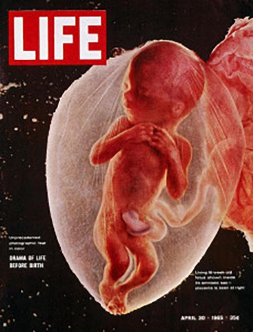
Exhibition Resources
Exhibition Dates:
January 2–April 12, 2019
Location:
Special Collections Research Center Exhibition Gallery,
1100 East 57th Street, Chicago, IL 60637
This exhibition has been featured in:
KNOW: A Journal on the Formation of Knowledge
‘The Fetus in Utero: From Mystery to Social Media’
Chicago Tribune
‘”The Fetus in Utero” at U. of C. reveals mysteries of the womb’
The Chicago Maroon
‘Embryonic Enigmas and Fetal Fantasies at Special Collections’
Formations
‘How did the Fetal Ultrasound Become Such an Iconic Image?’
Acknowledgments
The curators would like to thank all those who helped make this exhibition possible, including Special Collections staff, as well as our students and colleagues. We extend our thanks to the following individuals for their insights, contributions, and hard work in making this exhibition come together: Jacques Abramowicz, Julie Chor, Mike Kenny, Rachel Rosenberg, Mindy Schwartz, and the University of Chicago students who generously provided captions—Elizabeth Crowdus, Madeline de Figueiredo, Anya Dunaif, Chloe Hall, Anna Kheyfets, Amber Olson, and Antonia Willnow. We are grateful to the Stevanovich Institute on the Formation of Knowledge (SIFK) for their generous support, with special thanks to Shadi Bartsch-Zimmer, Macol Cerda, Richard Ruano, and Jessica Velazquez. The Polsky Center Fab Lab deserves a special thank you for creating the 3D printed fetus. The curators would like to extend additional gratitude to Teodora Szasz, Ashleigh Cassemere-Stanfield, and the Research Computing Center for making this virtual exhibit possible. Finally, we extend our gratitude to the fantastic Special Collections staff, including Catherine Uecker, Daniel Meyer, and especially Patti Gibbons, for their support at every stage in developing and executing this exhibition. We could not have done it without you.


